| Citation: |
JIA Muyuan, LI Ze, LIU Yuyang, LIU Jialin, ZHENG Xiaoque, BAI Yunjuan, CHEN Ling. Progress in Multidisciplinary Diagnosis and Treatment of Familial Brain Tumors[J]. Cancer Research on Prevention and Treatment, 2022, 49(6): 514-521. DOI: 10.3971/j.issn.1000-8578.2022.21.1511

|
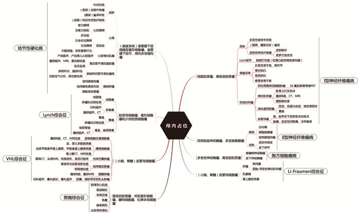
The tumors of central nervous system refer to a group of benign and malignant diseases originating from tissues or structures within the central nervous system. Common tumors of central nervous system are sporadic, but a few have familial onset. Compared with sporadic brain tumors, the clinical symptoms, diagnostic ideas and follow-up review plans of familial brain tumors are more complicated. The multidisciplinary diagnosis and treatment (MDT) mode usually refers to a treatment mode in which a case involving multiple organs and systems is discussed, and the best treatment plan is formulated for the patient based on the comprehensive opinions of various disciplines. Because familial brain tumors often involve multiple organs, multiple disciplines and multiple systems, and their low incidence leads to less clinical experience for neurosurgeons, the MDT model is more conducive to efficient diagnosis, treatment and management of familial brain tumors. This review elaborates on the neurosurgeon-led MDT model, and introduces the latest research on the epidemiology, genetic characteristics, clinical manifestations, diagnostic ideas and multidisciplinary management of familial brain tumors.
Competing interests: The authors declare that they have no competing interests.
| [1] |
Evans DG, Howard E, Giblin C, et al. Birth incidence and prevalence of tumor-prone syndromes: estimates from a UK family genetic register service[J]. Am J Med Genet A, 2010, 152A(2): 327-332. doi: 10.1002/ajmg.a.33139
|
| [2] |
Kehrer-Sawatzki H, Cooper DN. Challenges in the diagnosis of neurofibromatosis type 1 (NF1) in young children facilitated by means of revised diagnostic criteria including genetic testing for pathogenic NF1 gene variants[J]. Hum Genet, 2021, 141(2): 177-191.
|
| [3] |
Lloyd SK, Evans DG. Neurofibromatosis type 2 (NF2): diagnosis and management[J]. Handb Clin Neurol, 2013, 115: 957-967.
|
| [4] |
Farouk SS, Walsh MF, Karajannis MA. Genetic syndromes predisposing to pediatric brain tumors[J]. Neurooncol Pract, 2021, 8(4): 375-390.
|
| [5] |
Tamura R. Current Understanding of Neurofibromatosis Type 1, 2, and Schwannomatosis[J]. Int J Mol Sci, 2021, 22(11): 5850. doi: 10.3390/ijms22115850
|
| [6] |
Merker VL, Esparza S, Smith MJ, et al. Clinical features of schwannomatosis: a retrospective analysis of 87 patients[J]. Oncologist, 2012, 17(10): 1317-1322. doi: 10.1634/theoncologist.2012-0162
|
| [7] |
Carter JM, O'Hara C, Dundas G, et al. Epithelioid malignant peripheral nerve sheath tumor arising in a schwannoma, in a patient with "neuroblastoma-like" schwannomatosis and a novel germline SMARCB1 mutation[J]. Am J Surg Pathol, 2012, 36(1): 154-160. doi: 10.1097/PAS.0b013e3182380802
|
| [8] |
Bougeard G, Renaux-Petel M, Flaman JM, et al. Revisiting Li-Fraumeni Syndrome From TP53 Mutation Carriers[J]. J Clin Oncol, 2015, 33(21): 2345-2352. doi: 10.1200/JCO.2014.59.5728
|
| [9] |
Kumamoto T, Yamazaki F, Nakano Y, et al. Medical guidelines for Li-Fraumeni syndrome 2019, version 1.1[J]. Int J Clin Oncol, 2021, 26(12): 2161-2178. doi: 10.1007/s10147-021-02011-w
|
| [10] |
Northrup H, Krueger DA. Tuberous sclerosis complex diagnostic criteria update: recommendations of the 2012 Iinternational Tuberous Sclerosis Complex Consensus Conference[J]. Pediatr Neurol, 2013, 49(4): 243-254. doi: 10.1016/j.pediatrneurol.2013.08.001
|
| [11] |
Yates JR, Maclean C, Higgins JN, et al. The Tuberous Sclerosis 2000 Study: presentation, initial assessments and implications for diagnosis and management[J]. Arch Dis Child, 2011, 96(11): 1020-1025. doi: 10.1136/adc.2011.211995
|
| [12] |
Win AK, Jenkins MA, Dowty JG, et al. Prevalence and Penetrance of Major Genes and Polygenes for Colorectal Cancer[J]. Cancer Epidemiol Biomarkers Prev, 2017, 26(3): 404-412. doi: 10.1158/1055-9965.EPI-16-0693
|
| [13] |
Gong Y, Wang D, Wang W. Biostatistics of VHL-Gene Transfection in the Health Informatics Analysis of Renal Cell Carcinoma[J]. Comput Math Methods Med, 2022, 2022: 5297580.
|
| [14] |
Maher ER, Yates JR, Harries R, et al. Clinical features and natural history of von Hippel-Lindau disease[J]. Q J Med, 1990, 77(283): 1151-1163.
|
| [15] |
Romano AA, Allanson JE, Dahlgren J, et al. Noonan syndrome: clinical features, diagnosis, and management guidelines[J]. Pediatrics, 2010, 126(4): 746-759. doi: 10.1542/peds.2009-3207
|
| [16] |
Evans DG, Bowers NL, Tobi S, et al. Schwannomatosis: a genetic and epidemiological study[J]. J Neurol Neurosurg Psychiatry, 2018, 89(11): 1215-1219. doi: 10.1136/jnnp-2018-318538
|
| [17] |
Dutzmann CM, Vogel J, Kratz CP, et al. Update on Li-Fraumeni syndrome][J]. Pathologe, 2019, 40(6): 592-599. doi: 10.1007/s00292-019-00657-y
|
| [18] |
Deiller C, Van-Gils J, Zordan C, et al. Coexistence of schwannomatosis and glioblastoma in two families[J]. Eur J Med Genet, 2019, 62(8): 103680. doi: 10.1016/j.ejmg.2019.103680
|
| [19] |
Mai PL, Khincha PP, Loud JT, et al. Prevalence of Cancer at Baseline Screening in the National Cancer Institute Li-Fraumeni Syndrome Cohort[J]. JAMA Oncol, 2017, 3(12): 1640-1645. doi: 10.1001/jamaoncol.2017.1350
|
| [20] |
Hallett L, Foster T, Liu Z, et al. Burden of disease and unmet needs in tuberous sclerosis complex with neurological manifestations: systematic review[J]. Curr Med Res Opin, 2011, 27(8): 1571-1583. doi: 10.1185/03007995.2011.586687
|
| [21] |
Therkildsen C, Ladelund S, Rambech E, et al. Glioblastomas, astrocytomas and oligodendrogliomas linked to Lynch syndrome[J]. Eur J Neurol, 2015, 22(4): 717-724. doi: 10.1111/ene.12647
|
| [22] |
Khattab A, Monga DK. Turcot Syndrome[M]. StatPearls. Treasure Island (FL); StatPearls Publishing, 2021.
|
| [23] |
Bhambhani V, Muenke M. Noonan syndrome[J]. Am Fam Physician, 2014, 89(1): 37-43.
|
| [24] |
Dare AJ, Gupta AA, Thipphavong S, et al. Abdominal neoplastic manifestations of neurofibromatosis type 1[J]. Neurooncol Adv, 2020, 2(Suppl 1): i124-i133.
|
| [25] |
Suarez-Kelly LP, Yu L, Kline D, et al. Increased breast cancer risk in women with neurofibromatosis type 1: a meta-analysis and systematic review of the literature [J]. Hered Cancer Clin Pract, 2019, 17: 12. doi: 10.1186/s13053-019-0110-z
|
| [26] |
Castellanos E, Plana A, Carrato C, et al. Early Genetic Diagnosis of Neurofibromatosis Type 2 From Skin Plaque Plexiform Schwannomas in Childhood[J]. JAMA Dermatol, 2018, 154(3): 341-346. doi: 10.1001/jamadermatol.2017.5464
|
| [27] |
Plotkin SR, Bredella MA, Cai W, et al. Quantitative assessment of whole-body tumor burden in adult patients with neurofibromatosis[J]. PLoS One, 2012, 7(4): e35711. doi: 10.1371/journal.pone.0035711
|
| [28] |
Uysal SP, Şahin M. Tuberous sclerosis: a review of the past, present, andfuture[J]. Turk J Med Sci, 2020, 50(Si-2): 1665-1676.
|
| [29] |
Nair N, Chakraborty R, Mahajan Z, et al. Renal Manifestations of Tuberous Sclerosis Complex[J]. J Kidney Cancer VHL, 2020, 7(3): 5-19. doi: 10.15586/jkcvhl.2020.131
|
| [30] |
Taveira-DaSilva AM, Moss J. Epidemiology, pathogenesis and diagnosis of Lymphangioleiomyomatosis[J]. Exp Opin Orphan Drugs, 2016, 4(4): 369-378. doi: 10.1517/21678707.2016.1148597
|
| [31] |
Win AK, Lindor NM, Young JP, et al. Risks of primary extracolonic cancers following colorectal cancer in lynch syndrome[J]. J Nat Cancer Inst, 2012, 104(18): 1363-1372. doi: 10.1093/jnci/djs351
|
| [32] |
Verlinsky Y, Rechitsky S, Verlinsky O, et al. Preimplantation diagnosis for neurofibromatosis[J]. Reprod Biomed Online, 2002, 4(3): 218-222. doi: 10.1016/S1472-6483(10)61809-3
|
| [33] |
中国Ⅰ型神经纤维瘤病多中心治疗协作组, 全国整形外科多中心研究平台. Ⅰ型神经纤维瘤病临床诊疗专家共识(2021版)[J]. 中国修复重建外科杂志, 2021, 35(11): 1384-1395.
National Multi-Center Treatment Collaboration Group For Neurofibromatosis Type, National Multi-Center Research Platform For Plastic And Reconstructive Surgery, Reconstructive S. Expert consensus on diagnosis and management of neurofibromatosis type 1(2021 edition)[J]. Zhongguo Xiu Fu Chong Jian Wai Ke Za Zhi, 2021, 35(11): 1384-1395.
|
| [34] |
Sharif S, Ferner R, Birch JM, et al. Second primary tumors in neurofibromatosis 1 patients treated for optic glioma: substantial risks after radiotherapy[J]. J Clin Oncol, 2006, 24(16): 2570-2575. doi: 10.1200/JCO.2005.03.8349
|
| [35] |
Stewart DR, Sloan JL, Yao L, et al. Diagnosis, management and complications of glomus tumours of the digits in neurofibromatosis type 1[J]. J Med Genet, 2010, 47(8): 525-532. doi: 10.1136/jmg.2009.073965
|
| [36] |
Evans DGR, Salvador H, Chang VY, et al. Cancer and Central Nervous System Tumor Surveillance in Pediatric Neurofibromatosis 2 and Related Disorders[J]. Clin Cancer Res, 2017, 23(12): e54-e61. doi: 10.1158/1078-0432.CCR-17-0590
|
| [37] |
Asthagiri AR, Parry DM, Butman JA, et al. Neurofibromatosis type 2[J]. Lancet, 2009, 373(9679): 1974-1986. doi: 10.1016/S0140-6736(09)60259-2
|
| [38] |
Le AN, Harton J, Desai H, et al. Frequency of radiation-induced malignancies post-adjuvant radiotherapy for breast cancer in patients with Li-Fraumeni syndrome [J]. Breast Cancer Res Treat, 2020, 181(1): 181-188. doi: 10.1007/s10549-020-05612-7
|
| [39] |
Auranen A, Joutsiniemi T. A systematic review of gynecological cancer surveillance in women belonging to hereditary nonpolyposis colorectal cancer (Lynch syndrome) families[J]. Acta Obstet Gynecol Scand, 2011, 90(5): 437-444. doi: 10.1111/j.1600-0412.2011.01091.x
|
| [40] |
Syngal S, Brand RE, Church JM, et al. ACG clinical guideline: Genetic testing and management of hereditary gastrointestinal cancer syndromes[J]. Am J Gastroenterol, 2015, 110(2): 223-263. doi: 10.1038/ajg.2014.435
|
| [41] |
Tyburczy ME, Wang JA, Li S, et al. Sun exposure causes somatic second-hit mutations and angiofibroma development in tuberous sclerosis complex[J]. Hum Mol Genet, 2014, 23(8): 2023-2029. doi: 10.1093/hmg/ddt597
|
| [42] |
Gupta A, de Bruyn G, Tousseyn S, et al. Epilepsy and Neurodevelopmental Comorbidities in Tuberous Sclerosis Complex: A Natural History Study[J]. Pediatr Neurol, 2020, 106: 10-16. doi: 10.1016/j.pediatrneurol.2019.12.016
|
| [43] |
Giordano F, Moscheo C, Lenge M, et al. Neurosurgical treatment of subependymal giant cell astrocytomas in tuberous sclerosis complex: a series of 44 surgical procedures in 31 patients[J]. Childs Nerv Syst, 2020, 36(5): 951-960. doi: 10.1007/s00381-019-04449-w
|
| [44] |
Rednam SP, Erez A, Druker H, et al. Von Hippel-Lindau and Hereditary Pheochromocytoma/Paraganglioma Syndromes: Clinical Features, Genetics, and Surveillance Recommendations in Childhood[J]. Clin Cancer Res, 2017, 23(12): e68-e75. doi: 10.1158/1078-0432.CCR-17-0547
|
| [45] |
Frantzen C, Kruizinga RC, van Asselt SJ, et al. Pregnancy-related hemangioblastoma progression and complications in von Hippel-Lindau disease[J]. Neurology, 2012, 79(8): 793-796. doi: 10.1212/WNL.0b013e3182661f3c
|
| [46] |
Ordookhanian C, Kaloostian PE, Ghostine SS, et al. Management Strategiesand Outcomes for VHL-related Craniospinal Hemangioblastomas[J]. J kidney Cancer VHL, 2017, 4(3): 37-44. doi: 10.15586/jkcvhl.2017.90
|
| [47] |
李静, 肖静, 梁建宏. 视盘型视网膜毛细血管瘤的临床分析[J]. 中华眼科杂志, 2019, 55(8): 609-615. doi: 10.3760/cma.j.issn.0412-4081.2019.08.011
Li J, Xiao J, Liang JH. Clinical analysis of juxtapapillary retinal capillary hemangioma[J]. Zhonghua Yan Ke Za Zhi, 2019, 55(8): 609-615. doi: 10.3760/cma.j.issn.0412-4081.2019.08.011
|
| [48] |
Fang F, Ding L, He Q, et al. Preoperative Management of Pheochromocytoma and Paraganglioma[J]. Front Endocrinol (Lausanne), 2020, 11: 586795. doi: 10.3389/fendo.2020.586795
|
| [49] |
Gravholt CH, Viuff MH, Brun S, et al. Turner syndrome: mechanisms and management[J]. Nat Rev Endocrinol, 2019, 15(10): 601-614. doi: 10.1038/s41574-019-0224-4
|
| [1] | ZENG Lingcheng, LI Hua, CHEN Rudong, YANG Hongkuan, CHEN Jian, YU Jiasheng. Molecular Pathological Risk Grade Evaluates Biological Behavior and Prognosis of Patients with WHO Grade 1 Meningiomas[J]. Cancer Research on Prevention and Treatment, 2024, 51(6): 455-461. DOI: 10.3971/j.issn.1000-8578.2024.23.1342 |
| [2] | HAN Dong, ZHANG Xirong, JIA Yongjun, REN Ge, LYU Ruihua, SHI Linna, HE Taiping. A Neural Network Model Based on Enhanced CT for Distinguishing ISUP Grade of Clear Cell Renal Cell Carcinoma[J]. Cancer Research on Prevention and Treatment, 2021, 48(1): 55-59. DOI: 10.3971/j.issn.1000-8578.2021.20.0440 |
| [3] | SONG Jinling, LI Zhongwu, WEI Maomao, ZHOU Ni'na, YANG Zhi, WANG Xuejuan. Relation Between Metabolic Parameters of 18F-FDG PET/CT and Clinicopathological Features of Colorectal Cancer Patients[J]. Cancer Research on Prevention and Treatment, 2019, 46(11): 1006-1012. DOI: 10.3971/j.issn.1000-8578.2019.19.0665 |
| [4] | ZHOU Ni'na, ZHU Hua, YANG Zhi. Progress in PET/CT Molecular Imaging Targeting HER2-positive Tumour[J]. Cancer Research on Prevention and Treatment, 2019, 46(4): 376-381. DOI: 10.3971/j.issn.1000-8578.2019.18.1016 |
| [5] | ZHOU Ni'na, YU Jiangyuan, ZHU Hua, ZHAO Wei, YANG Zhi. PET/CT Imaging Characteristics of Abdominal Fibroblastic/Myofibroblastic Tumors and Its Application Value[J]. Cancer Research on Prevention and Treatment, 2019, 46(3): 243-247. DOI: 10.3971/j.issn.1000-8578.2019.18.1817 |
| [6] | LU Shengnan, FENG Yanlin, LI Wen, WANG Ying, XIAN Weijun. Correlation Between 18F-FDG PET/CT SUVmax and Clinicopathological Features, Neoadjuvant Chemotherapy Response in Invasive Ductal Breast Carcinoma Patients[J]. Cancer Research on Prevention and Treatment, 2019, 46(2): 144-148. DOI: 10.3971/j.issn.1000-8578.2019.18.1230 |
| [7] | ZHANG Qiang, SUN Lijiang, ZHANG Guiming, QI Jun, YANG Zhigang. Influence of Periprostatic Adiposity Measurement Parameters on Preoperative Staging and Grading of Prostate Cancer[J]. Cancer Research on Prevention and Treatment, 2018, 45(7): 488-491. DOI: 10.3971/j.issn.1000-8578.2018.17.1382 |
| [8] | TIAN Jinhui, LI Jinlong, GE Long, WANG Jing, ZHANG Mingxia, SHI Fangyu, WANG Xiaohu, ZHANG Hong. Diagnostic Value of PET/CT for Metastatic Lymph Nodes in Cervical Cancer Patients: A Meta-analysis[J]. Cancer Research on Prevention and Treatment, 2015, 42(03): 270-276. DOI: 10.3971/j.issn.1000-8578.2015.03.013 |
| [9] | FengYanlin, Lu Shengnan, Yang Jie, Xian Weijun. Valuation of 18F-FDG PET/CT in Predicting Pathological Reaction of Neoadjuvant Chemotherapy with Breast Cancer[J]. Cancer Research on Prevention and Treatment, 2012, 39(10): 1224-1227. DOI: 10.3971/j.issn.1000-8578.2012.10.015 |
| [10] | SHU Qing, ZHANG Su zhen, WEI Ya ning, MA Qun feng. Methylation of Androgen receptor Gene and the Grade of Esophageal Cancer[J]. Cancer Research on Prevention and Treatment, 2004, 31(11): 688-690. DOI: 10.3971/j.issn.1000-8578.2081 |
Figures(1) / Tables(2)

This work is licensed under a Creative Commons Attribution 3.0 License.
Copyright © Editorial Department of Cancer Prevention Research 鄂公网安备 42011102005013号 鄂ICP备2022015867号
Supported by: Beijing Renhe Information Technology Co., Ltd. 
 DownLoad: