| Citation: |
ZHOU Jieli, WU Linjuan, ZHANG Pengtian, Peng Yanxia, HAN Dong. Prediction of pN Staging of Papillary Thyroid Carcinoma Using Ultrasonography Radiomics and Deep Neural Networks[J]. Cancer Research on Prevention and Treatment, 2025, 52(2): 151-155. DOI: 10.3971/j.issn.1000-8578.2025.24.0617

|
To assess the accuracy of pN staging prediction in papillary thyroid carcinoma (PTC) using ultrasound radiomics and deep neural networks (DNN).
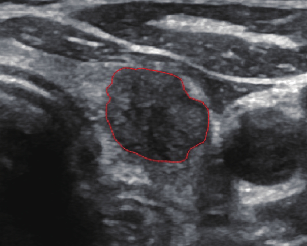
A retrospective analysis was conducted on 375 patients with pathologically confirmed PTC, comprising 261 cases in the training set and 114 in the test set. Staging was categorized as pN0 (no cervical lymph node metastasis), pN1a (central neck lymph node metastasis), and pN1b (lateral neck lymph node metastasis). An ultrasound physician manually segmented the regions of interest (ROIs) for PTC, extracting
A total of 153 patients were in the pN0 stage, 131 patients in the pN1a stage, and 91 patients in the pN1b stage. LASSO regression selected 15 radiomic features for each PTC. The optimal DNN model, constructed using these 15 features, achieved accuracies of 85.82% on the training set and 81.57% on the test set.
Ultrasound radiomics of PTC demonstrates high accuracy in predicting pN staging and shows potential for automating N staging in patients.
Competing interests: The authors declare that they have no competing interests.
| [1] |
Chow SM, Law SC, Chan JK, et al. Papillary microcarcinoma of the thyroid-Prognostic significance of lymph node metastasis and multifocality[J]. Cancer, 2003, 98(1): 31-40. doi: 10.1002/cncr.11442
|
| [2] |
Lim YC, Choi EC, Yoon YH, et al. Central lymph node metastases in unilateral papillary thyroid microcarcinoma[J]. Br J Surg, 2009, 96(3): 253-257. doi: 10.1002/bjs.6484
|
| [3] |
Shaha AR. Prognostic factors in papillary thyroid carcinoma and implications of large nodal metastasis[J]. Surgery, 2004, 135(2): 237-239. doi: 10.1016/j.surg.2003.08.023
|
| [4] |
Haugen BR, Alexander EK, Bible KC, et al. 2015 American thyroid association management guidelines for adult patients with thyroid nodules and differentiated thyroid cancer: The American thyroid association guidelines task force on thyroid nodules and differentiated thyroid cancer[J]. Thyroid, 2016, 26(1): 1-133. doi: 10.1089/thy.2015.0020
|
| [5] |
Podnos YD, Smith D, Wagman LD, et al. The implication of lymph node metastasis on survival in patients with well-differentiated thyroid cancer[J]. Am Surg, 2005, 71(9): 731-734. doi: 10.1177/000313480507100907
|
| [6] |
Zaydfudim V, Feurer ID, Griffin MR, et al. The impact of lymph node involvement on survival in patients with papillary and follicular thyroid carcinoma[J]. Surgery, 2008, 144(6): 1070-1078. doi: 10.1016/j.surg.2008.08.034
|
| [7] |
Liu FH, Kuo SF, Hsueh C, et al. Postoperative recurrence of papillary thyroid carcinoma with lymph node metastasis[J]. J Surg Oncol, 2015, 112(2): 149-154. doi: 10.1002/jso.23967
|
| [8] |
Luo XY, Chen AM, Zhou Y, et al. Analysis of risk factors for postoperative recurrence of thyroid cancer[J]. J BUON, 2019, 24(2): 813-818.
|
| [9] |
Moo TA, McGill J, Allendorf J, et al. Impact of prophylactic central neck lymph node dissection on early recurrence in papillary thyroid carcinoma[J]. World J Surg, 2010, 34(6): 1187-1191. doi: 10.1007/s00268-010-0418-3
|
| [10] |
Goropoulos A, Karamoshos K, Christodoulou A, et al. Value of the cervical compartments in the surgical treatment of papillary thyroid carcinoma[J]. World J Surg, 2004, 28(12): 1275-1281. doi: 10.1007/s00268-004-7643-6
|
| [11] |
Gemsenjäger E, Perren A, Seifert B, et al. Lymph node surgery in papillary thyroid carcinoma[J]. J Am Coll Surg, 2003, 197(2): 182-190. doi: 10.1016/S1072-7515(03)00421-6
|
| [12] |
American thyroid association (ATA) guidelines taskforce on thyroid nodules and differentiated thyroid cancer, Cooper DS, Doherty GM, et al. Revised American thyroid association management guidelines for patients with thyroid nodules and differentiated thyroid cancer[J]. Thyroid, 2009, 19(11): 1167-1214. doi: 10.1089/thy.2009.0110
|
| [13] |
Randolph GW, Duh QY, Heller KS, et al. The prognostic significance of nodal metastases from papillary thyroid carcinoma can be stratified based on the size and number of metastatic lymph nodes, as well as the presence of extranodal extension[J]. Thyroid, 2012, 22(11): 1144-1152. doi: 10.1089/thy.2012.0043
|
| [14] |
Kim E, Park JS, Son KR, et al. Preoperative diagnosis of cervical metastatic lymph nodes in papillary thyroid carcinoma: comparison of ultrasound, computed tomography, and combined ultrasound with computed tomography[J]. Thyroid, 2008, 18(4): 411-418. doi: 10.1089/thy.2007.0269
|
| [15] |
Jeong HS, Baek CH, Son YI, et al. Integrated 18F-FDG PET/CT for the initial evaluation of cervical node level of patients with papillary thyroid carcinoma: comparison with ultrasound and contrast-enhanced CT[J]. Clin Endocrinol (Oxf), 2006, 65(3): 402-407. doi: 10.1111/j.1365-2265.2006.02612.x
|
| [16] |
hoi JS, Kim J, Kwak JY, et al. Preoperative staging of papillary thyroid carcinoma: comparison of ultrasound imaging and CT[J]. Am J Roentgenol, 2009, 193(3): 871-878. doi: 10.2214/AJR.09.2386
|
| [17] |
Gillies RJ, Kinahan PE, Hricak H. Radiomics: images are more than pictures, they are data[J]. Radiology, 2016, 278(2): 563-577. doi: 10.1148/radiol.2015151169
|
| [18] |
Paul AY, Joseph P, Heather CH, et al. User-guided 3D active contour segmentation of anatomical structures: Significantly improved efficiency and reliability[J]. Neuroimage, 2006, 31(3): 1116-1128. doi: 10.1016/j.neuroimage.2006.01.015
|
| [19] |
Zwanenburg A, Vallières M, Abdalah MA, et al. The image biomarker standardization initiative: standardized quantitative radiomics for high-throughput image-based phenotyping[J]. Radiology, 2020, 295(2): 328-338. doi: 10.1148/radiol.2020191145
|
| [20] |
Kim SY, Lee E, Nam SJ, et al. Ultrasound texture analysis: association with lymph node metastasis of papillary thyroid microcarcinoma[J]. PLoS One, 2017, 12(4): e0176103. doi: 10.1371/journal.pone.0176103
|
| [21] |
Park VY, Han K, Kim HJ, et al. Radiomics signature for prediction of lateral lymph node metastasis in conventional papillary thyroid carcinoma[J]. PLoS One, 2020, 15(1): e0227315. doi: 10.1371/journal.pone.0227315
|
| [22] |
Liu T, Zhou S, Yu J, et al. Prediction of lymph node metastasis in patients with papillary thyroid carcinoma: a radiomics method based on preoperative ultrasound images[J]. Technol Cancer Res Treat, 2019, 18: 1533033819831713.
|
| [23] |
Liu T, Ge X, Yu J, et al. Comparison of the application of B-mode and strain elastography ultrasound in the estimation of lymph node metastasis of papillary thyroid carcinoma based on a radiomics approach[J]. Int J Comput Assist Radiol Surg, 2018, 13(10): 1617-1627. doi: 10.1007/s11548-018-1796-5
|
| [24] |
黄云霞, 周瑾, 刘桐桐, 等. 超声影像组学与传统影像模式对甲状腺乳头状癌颈部中央区淋巴结转移的诊断价值比较[J]. 中华超声影像学杂志, 2019, 28(10): 882-887. [Huang YX, Zhou J, Liu TT, et al. Comparison of ultrasound radiomics with conventional imaging models: diagnosis of central cervical lymph node metastasis in papillary thyroid carcinoma[J]. Zhonghua Chao Sheng Ying Xiang Xue Za Zhi, 2019, 28(10): 882-887.] doi: 10.3760/cma.j.issn.1004-4477.2019.10.011
Huang YX, Zhou J, Liu TT, et al. Comparison of ultrasound radiomics with conventional imaging models: diagnosis of central cervical lymph node metastasis in papillary thyroid carcinoma[J]. Zhonghua Chao Sheng Ying Xiang Xue Za Zhi, 2019, 28(10): 882-887. doi: 10.3760/cma.j.issn.1004-4477.2019.10.011
|
| [25] |
Jiang M, Li C, Tang S, et al. Nomogram based on shear-wave elastography radiomics can improve preoperative cervical lymph node staging for papillary thyroid carcinoma[J]. Thyroid, 2020, 30(6): 885-897. doi: 10.1089/thy.2019.0780
|
| [26] |
Xiao Q, Zhu W, Tang H, et al. Ultrasound radiomics in the prediction of microvascular invasion in hepatocellular carcinoma: A systematic review and meta-analysis[J]. Heliyon, 2023, 9(6): e16997. doi: 10.1016/j.heliyon.2023.e16997
|
| [27] |
葛晓燕, 韩红娟, 罗艳虹, 等. 基于神经影像数据的阿尔茨海默病多分类诊断模型的研究进展与挑战[J]. 郑州大学学报(医学版), 2020, 55(1): 1-7. [Ge XY, Han HJ, Luo YH, et al. Research progress and challenges of multi class diagnosis model for Alzheimer's disease based on neuroimaging data[J]. Zhengzhou Da Xue Xue Bao (Yi Xue Ban), 2020, 55(1): 1-7.]
Ge XY, Han HJ, Luo YH, et al. Research progress and challenges of multi class diagnosis model for Alzheimer's disease based on neuroimaging data[J]. Zhengzhou Da Xue Xue Bao (Yi Xue Ban), 2020, 55(1): 1-7.
|
Figures(3) / Tables(1)

This work is licensed under a Creative Commons Attribution 3.0 License.
Copyright © Editorial Department of Cancer Prevention Research 鄂公网安备 42011102005013号 鄂ICP备2022015867号
Supported by: Beijing Renhe Information Technology Co., Ltd. 
 DownLoad: