| Citation: |
ZENG Chunyuan, CHENG Yong, XU Hao. Diagnostic Value of 18F-labeled PSMA PET/CT for Regional Lymph Node Metastasis in Prostate Cancer: A Meta-analysis[J]. Cancer Research on Prevention and Treatment, 2022, 49(2): 141-147. DOI: 10.3971/j.issn.1000-8578.2022.21.0752

|
To evaluate the diagnostic value of 18F-labeled PSMA PET/CT for regional lymph node metastasis in prostate cancer.
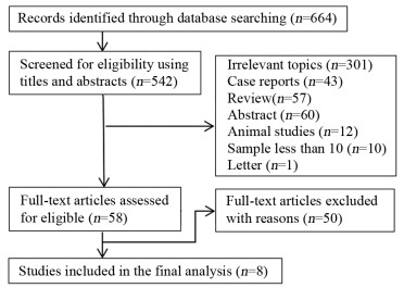
We searched PubMed, Embase, Cochrane Library, Web of Science, CNKI, VIP and Wanfang database from January 1, 2000 to May 31, 2021 for the studies about the diagnosis of 18F-labeled PSMA PET/CT for regional lymph node metastasis and staging in prostate cancer. Two investigators screened literature, extracted relevant data and assessed the quality of the literature independently. The meta-analysis was performed using Meta-disc 1.4 and Stata 16.0 software.
A total of 8 studies were finally included for the analysis, consisting of 754 prostate cancer patients and 2101 lymph nodes. The results of this meta-analysis showed pooled sensitivity, pooled specificity, pooled positive likelihood ratio, pooled negative likelihood ratio and diagnostic ratio were 0.82 (95%CI: 0.61-0.93), 0.98 (95%CI: 0.91-1.00), 45.7 (95%CI: 9.0-231.3), 0.18 (95%CI: 0.07-0.45) and 251 (95%CI: 34-1851), respectively. The area under the SROC curve was 0.97 (95%CI: 0.95-0.98).
18F-labeled PSMA PET/CT has a high diagnostic value for regional lymph node metastasis in prostate cancer.
Competing interests: The authors declare that they have no competing interests.
| [1] |
Siegel RL, Miller KD, Fuchs HE, et al. Cancer statistics, 2021[J]. CA Cancer J Clin, 2021, 71(1): 7-33. doi: 10.3322/caac.21654
|
| [2] |
Ieiri K, Shiota M, Kashiwagi E, et al. The prognosis and the impact of radiotherapy in clinically regional lymph node-positive prostate cancer: which patients are candidates for local therapy with radiation?[J]. Urol Oncol, 2020, 38(12): 931. e1-931. e7. doi: 10.1016/j.urolonc.2020.08.018
|
| [3] |
Hinsenveld FJ, Wit EMK, van Leeuwen PJ, et al. Prostate-specific membrane antigen PET/CT combined with sentinel node biopsy for primary lymph node staging in prostate cancer[J]. J Nucl Med, 2020, 61(4): 540-545. doi: 10.2967/jnumed.119.232199
|
| [4] |
Zhang Q, Zang S, Zhang C, et al. Comparison of 68Ga-PSMA-11 PET-CT with mpMRI for preoperative lymph node staging in patients with intermediate to high-risk prostate cancer[J]. J Transl Med, 2017, 15(1): 230. doi: 10.1186/s12967-017-1333-2
|
| [5] |
Maurer T, Gschwend JE, Rauscher I, et al. Diagnostic efficacy of 68Gallium-PSMA positron emission tomography compared to conventional imaging for lymph node staging of 130 consecutive patients with intermediate to high risk prostate cancer[J]. J Urol, 2016, 195(5): 1436-1443. doi: 10.1016/j.juro.2015.12.025
|
| [6] |
Kesch C, Kratochwil C, Mier W, et al. 68Ga or 18F for prostate cancer imaging?[J]. J Nucl Med, 2017, 58(5): 687-688. doi: 10.2967/jnumed.117.190157
|
| [7] |
Sanchez-Crespo A. Comparison of Gallium-68 and Fluorine-18 imaging characteristics in positron emission tomography[J]. Appl Radiat Isot, 2013, 76: 55-62. doi: 10.1016/j.apradiso.2012.06.034
|
| [8] |
Giesel FL, Hadaschik B, Cardinale J, et al. F-18 labelled PSMA-1007: biodistribution, radiation dosimetry and histopathological validation of tumor lesions in prostate cancer patients[J]. Eur J Nucl Med Mol Imaging, 2017, 44(4): 678-688. doi: 10.1007/s00259-016-3573-4
|
| [9] |
Jansen BHE, Bodar YJL, Zwezerijnen GLC, et al. Pelvic lymph-node staging with 18F-DCFPyL PET/CT prior to extended pelvic lymph-node dissection in primary prostate cancer-the SALT trial[J]. Eur J Nucl Med Mol Imaging, 2021, 48(2): 509-520. doi: 10.1007/s00259-020-04974-w
|
| [10] |
Gorin MA, Rowe SP, Patel HD, et al. Prostate specific membrane antigen targeted 18F-DCFPyL positron emission tomography/computerized tomography for the preoperative staging of high risk prostate cancer: results of a prospective, phase Ⅱ, single center study[J]. J Urol, 2018, 199(1): 126-132. doi: 10.1016/j.juro.2017.07.070
|
| [11] |
Malaspina S, Anttinen M, Taimen P, et al. Prospective comparison of 18F-PSMA-1007 PET/CT, whole-body MRI and CT in primary nodal staging of unfavourable intermediate- and high-risk prostate cancer[J]. Eur J Nucl Med Mol Imaging, 2021, 48(9): 2951-2959. doi: 10.1007/s00259-021-05296-1
|
| [12] |
Sprute K, Kramer V, Koerber SA, et al. Diagnostic accuracy of 18F-PSMA-1007 PET/CT imaging for lymph node staging of prostate carcinoma in primary and biochemical recurrence[J]. J Nucl Med, 2021, 62(2): 208-213. doi: 10.2967/jnumed.120.246363
|
| [13] |
Pienta KJ, Gorin MA, Rowe SP, et al. A phase 2/3 prospective multicenter study of the diagnostic accuracy of prostate specific membrane antigen PET/CT with 18F-DCFPyL in prostate cancer patients (OSPREY)[J]. J Urol, 2021, 206(1): 52-61. doi: 10.1097/JU.0000000000001698
|
| [14] |
Lindenberg L, Mena E, Turkbey B, et al. Evaluating biochemically recurrent prostate cancer: histologic validation of 18F-DCFPyL PET/CT with comparison to multiparametric MRI[J]. Radiology, 2020, 296(3): 564-572. doi: 10.1148/radiol.2020192018
|
| [15] |
刘亚超, 刘家金, 张晓军, 等. 18F-DCFPyL PET/CT术前诊断前列腺癌区域转移淋巴结[J]. 中国医学影像技术, 2020, 36(6): 868-872. https://www.cnki.com.cn/Article/CJFDTOTAL-ZYXX202006020.htm
Liu YC, Liu JJ, Zhang XJ, et al. 18F-DCFPyL PET/CT in pre-operative diagnosis of regional lymph node metastasis from prostate cancer[J]. Zhongguo Yi Xue Ying Xiang Ji Shu, 2020, 36 (6): 868-872. https://www.cnki.com.cn/Article/CJFDTOTAL-ZYXX202006020.htm
|
| [16] |
Whiting PF, Rutjes AW, Westwood ME, et al. QUADAS-2: a revised tool for the quality assessment of diagnostic accuracy studies[J]. Ann Intern Med, 2011, 155(8): 529-536. doi: 10.7326/0003-4819-155-8-201110180-00009
|
| [17] |
Nini A, Gandaglia G, Fossati N, et al. Patterns of clinical recurrence of node-positive prostate cancer and impact on long-term survival[J]. Eur Urol, 2015, 68(5): 777-784. doi: 10.1016/j.eururo.2015.04.035
|
| [18] |
Trabulsi EJ, Rumble RB, Jadvar H, et al. Optimum imaging strategies for advanced prostate cancer: ASCO guideline[J]. J Clin Oncol, 2020, 38(17): 1963-1996. doi: 10.1200/JCO.19.02757
|
| [19] |
Kim SJ, Lee SW, Ha HK. Diagnostic performance of radiolabeled prostate-specific membrane antigen positron emission tomography/computed tomography for primary lymph node staging in newly diagnosed intermediate to high-risk prostate cancer patients: a systematic review and meta-analysis[J]. Urol Int, 2019, 102(1): 27-36. doi: 10.1159/000493169
|
| [20] |
Tu X, Zhang C, Liu Z, et al. The role of 68Ga-PSMA positron emission tomography/computerized tomography for preoperative lymph node staging in intermediate/high risk patients with prostate cancer: a diagnostic meta-analysis[J]. Front Oncol, 2020, 10: 1365. doi: 10.3389/fonc.2020.01365
|
| [21] |
Dietlein F, Kobe C, Hohberg M, et al. Intraindividual comparison of 18F-PSMA-1007 with renally excreted PSMA ligands for PSMA PET imaging in patients with relapsed prostate cancer[J]. J Nucl Med, 2020, 61(5): 729-734. doi: 10.2967/jnumed.119.234898
|
| [22] |
Giesel FL, Knorr K, Spohn F, et al. Detection efficacy of 18F-PSMA-1007 PET/CT in 251 patients with biochemical recurrence of prostate cancer after radical prostatectomy[J]. J Nucl Med, 2019, 60(3): 362-368. doi: 10.2967/jnumed.118.212233
|
| [23] |
Gaur S, Mena E, Harmon SA, et al. Prospective evaluation of 18F-DCFPyL PET/CT in detection of high-risk localized prostate cancer: comparison with mpMRI[J]. AJR Am J Roentgenol, 2020, 215(3): 652-659. doi: 10.2214/AJR.19.22042
|
| [24] |
Wondergem M, van der Zant FM, Broos WAM, et al. 18F-DCFPyL PET/CT for primary staging in 160 high-risk prostate cancer patients; metastasis detection rate, influence on clinical management and preliminary results of treatment efficacy[J]. Eur J Nucl Med Mol Imaging, 2021, 48(2): 521-531. doi: 10.1007/s00259-020-04782-2
|
| [25] |
Hövels AM, Heesakkers RA, Adang EM, et al. The diagnostic accuracy of CT and MRI in the staging of pelvic lymph nodes in patients with prostate cancer: a meta-analysis[J]. Clin Radiol, 2008, 63(4): 387-395. doi: 10.1016/j.crad.2007.05.022
|
| 1. |
吴迎朝,朱仕奇,梁嘉谊,梁裕琪,陈柳汕,陈洁婷,左谦,陈前军. 逍遥散通过减轻海马神经炎症缓解乳腺癌性抑郁. 中草药. 2025(11): 3910-3919 .

|
Figures(5) / Tables(2)

This work is licensed under a Creative Commons Attribution 3.0 License.
Copyright © Editorial Department of Cancer Prevention Research 鄂公网安备 42011102005013号 鄂ICP备2022015867号
Supported by: Beijing Renhe Information Technology Co., Ltd. 
 DownLoad: