| Citation: |
ZHOU Ni'na, LI Nan, WANG Xuejuan, ZHU Hua, FAN Yang, YANG Zhi. PET/CT Manifestations of Different Pathological Subtypes of Retroperitoneal Liposarcoma[J]. Cancer Research on Prevention and Treatment, 2018, 45(5): 316-319. DOI: 10.3971/j.issn.1000-8578.2018.17.1628

|
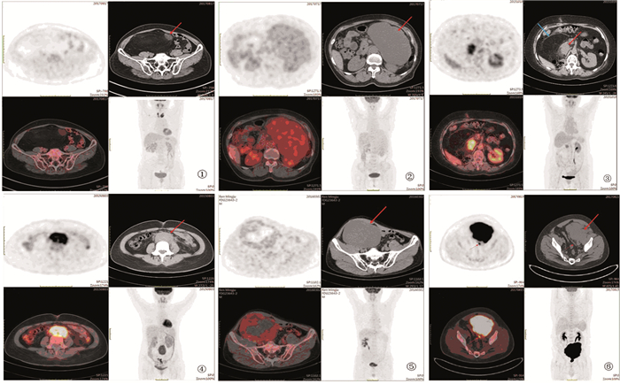
To analyze PET/CT imaging manifestations of different pathological subtypes of retroperitoneal liposarcoma, to explore the application value of PET/CT in retroperitoneal liposarcoma.
We included 48 patients diagnosed pathologically as retroperitoneal liposarcoma. According to the classification of WHO2013 soft tissue tumor and FNCCLE criterion, the patients were divided into three groups, G1 group included nine well-differentiated liposarcoma(WDLS), G2 group included seven myxoid liposarcoma (MLS), and G3 group included 29 dedifferentiated liposarcoma (DDLS), two pleomorphic liposarcoma(PLS), and one round cell liposarcoma(RCLS). PET/CT manifestations of different subtypes of retroperitoneal liposarcoma were analyzed. FDG metabolic degree and uniformity among three groups were compared by ANOVA test.
(1) WDLS showed fat density mass with septa and small nodules, with mild FDG uptake; MLS showed low density mass with mild to moderate FDG uptake, with or without fat; DDLS/PLS/RCLS showed soft tissue mass with high FDG uptake, with or without fat; (2)All lesions had local invasion, without lymph node metastasis; one recurrent DDLS had meditational metastasis; one RCLS had peritoneal metastasis; (3)The mean SUVmax G1 < G2 < G3 (2.6±1.6, 4.6±2.4, 9.8±9.3), there was significant difference between G1 and G3 groups(P < 0.05).
Different subtypes of retroperitoneal liposarcoma have different PET/CT manifestations. Preoperative PET/CT could accurately show the extent and metastasis of tumor and predict the pathological subtype, which could help the design of surgical scheme.
| [1] |
Taguchi S, Kume H, Fukuhara H, et al. Symptoms at diagnosis as independent prognostic factors in retroperi-toneal liposarcoma[J]. Mol Clin Oncol, 2016, 4(2): 255-60. doi: 10.3892/mco.2015.701
|
| [2] |
Vijay A, Ram L. Retroperitoneal liposarcoma: A comprehensive review[J]. Am J Clin Oncol, 2015, 38(2): 213-9. doi: 10.1097/COC.0b013e31829b5667
|
| [3] |
Fletchero CDM, Bridge JA, Hogendoorn PCW, et al. World Health Organization classification of soft tissue and bone tumours[M]. Lyon: IARCP Press, 2013.
|
| [4] |
林翠君, 李丽红, 黄春榆, 等.脂肪肉瘤的CT、MRI表现与病理学对照[J].中国CT和MRI杂志, 2015, 13(8): 108-11. http://med.wanfangdata.com.cn/Paper/Detail/PeriodicalPaper_zgcthmrizz201508033
Lin CJ, Li LH, Huang CY, et al. CT and MRI Manifestations of Liposarcoma with Pathologic Features[J]. Zhongguo CT He MRI Za Zhi, 2015, 13(8): 108-11. http://med.wanfangdata.com.cn/Paper/Detail/PeriodicalPaper_zgcthmrizz201508033
|
| [5] |
Dong M, Bi J, Liu X, et al. Significant partial response of metastatic intra-abdominal and pelvic round cell liposarcoma to a small-molecule VEGFR-2 tyrosine kinase inhibitor apatinib: A case report[J]. Medicine (Baltimore), 2016, 95(31): e4368. doi: 10.1097/MD.0000000000004368
|
| [6] |
Oh SD, Oh SJ, Suh BJ, et al. A Giant Retroperitoneal liposarcoma encasing the entire left kidney and adherent to adjacent structures: A case report[J]. Case Rep Oncol, 2016, 9(2): 368-72. doi: 10.1159/000447488
|
| [7] |
Sharma M, Mannan R, Bhasin TS, et al. Giant inflammatory variant of well differentiated liposarcoma: a case report of a rare entity[J]. J Clin Diagn Res, 2013, 7(8): 1720-1. http://europepmc.org/abstract/med/24086890
|
| [8] |
Morosi C, Stacchiotti A, Marchianò A, et al. Correlation between radiological assessment and histopathological diagnosis in retroperitoneal tumors:Analysis of 291 consecutive patients at a tertiary reference sarcoma center[J]. Eur J Surg Oncol, 2014, 40(12): 1662-70. doi: 10.1016/j.ejso.2014.10.005
|
| [9] |
Lee SY, Goh BK, Teo MC, et al. Retroperitoneal liposarcomas: the experience of a tertiary Asian center[J]. World J Surg Oncol, 2011, 9: 12. doi: 10.1186/1477-7819-9-12
|
| [10] |
Singer S, Antonescu CR, Riedel E, et al. Histologic subtype and margin of resection Predict pattern of recurrence and survival for retroperitoneal liposarcoma[J]. Ann Surg, 2003, 238(3): 358-70. http://europepmc.org/abstract/MED/14501502
|
| [11] |
Mansfield SA, Pollock RE, Grignol VP. Surgery for Abdominal Well-Differentiated Liposarcoma[J]. Curr Treat Options Oncol, 2018, 19(1): 1. doi: 10.1007/s11864-018-0520-6
|
| [12] |
Trans-Atlantic RPS Working Group. Management of recurrent retroperitoneal sarcoma (RPS) in the adult: A consensus approach from the trans-atlantic RPS working group[J]. Ann Surg Oncol, 2016, 23(11): 3531-40. doi: 10.1245/s10434-016-5336-7
|
| [13] |
Zeng X, Liu W, Wu X, et al. Clinicopathological characteristics and experience in the treatment of giant retroperitoneal liposarcoma: A case report and review of the literature[J]. Cancer Biol Ther, 2017, 18(9): 660-5. doi: 10.1080/15384047.2017.1345388
|
| [14] |
Stahl JM, Corso CD, Park HS, et al. The effect of microscopic margin status on survival in adult retroperitoneal soft tissue sarcomas[J]. Eur J Surg Oncol, 2017, 43(1): 168-74. doi: 10.1016/j.ejso.2016.05.031
|
| [15] |
Molina G, Hull MA, Chen YL, et al. Preoperative radiation therapy combined with radical surgical resection is associated with a lower rate of local recurrence when treating unifocal, primary retroperitoneal liposarcoma[J]. J Surg Oncol, 2016, 114(7): 814-20. doi: 10.1002/jso.24427
|

This work is licensed under a Creative Commons Attribution 3.0 License.
Copyright © Editorial Department of Cancer Prevention Research 鄂公网安备 42011102005013号 鄂ICP备2022015867号
Supported by: Beijing Renhe Information Technology Co., Ltd. 
 DownLoad: