| Citation: |
DUAN Baojun, BAI Jun, GUO Yanfeng, PU Yansong, MA Guodong. Established of Serum Diagnostic Model for Colorectal Cancer Patients Using MB-WCX and MALDI-TOF MS[J]. Cancer Research on Prevention and Treatment, 2018, 45(6): 386-390. DOI: 10.3971/j.issn.1000-8578.2018.17.1276

|
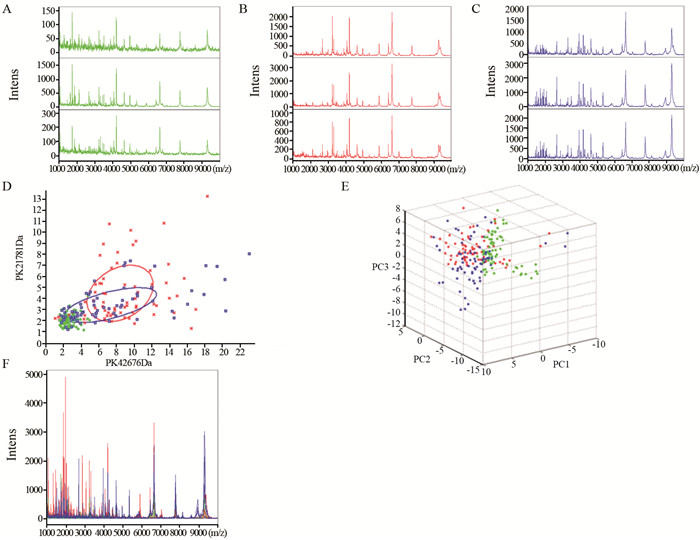
Serum protein expression profiling was examined using magnetic bead-based matrix-assisted laser desorption/ionization time-of-flight (MALDI-TOF-MS) to establish a serum proteomic diagnostic model for colorectal cancer.
Serum samples of normal control (CRTL, n=72), colorectal cancer (pre-operation CRC, n=72, and post-operation CRC, n=72) were collected from 2014-9-1 to 2016-9-1. Peptidome of all samples were extracted by magnetic-bead-based weak cation-exchange chromatography (MB-WCX) and detected by calibrated Autoflex Ⅲ MALDI-TOF-MS. Peptide mass fingerprinting were analyzed by ClinProtTools 2.0 software, and the differentially-expressioned peptides were further identified using LC-ESI-MS/MS.
MALDI-TOF-MS identified 80 peaks (m/z), in which 12 peaks showed significant differences among CRTL, pre-operation and post-operation CRC patients (P < 0.01). 9 peaks were up-regulated and 3 peaks were down-regulated in CRC compared with CRTL, and these peaks showed a tendency to CRTL after operation. Based on the GA model, CRC patients could be discriminated from CRTL with 99.31% sensitivity and 96.49% specificity. Moreover, 3 peaks (m/z: 2663.36, m/z: 4793.17 and m/z: 5343.48) of the GA model were identified as protein FGA, SETD7 and MUC5AC respectively.
The serum proteomic diagnostic model could accurately distinguish between CRTL and CRC, but it needs further research.
| [1] |
Favoriti P, Carbone G, Greco M, et al. Worldwide burden of colorectal cancer: a review[J]. Updates Surg, 2016, 68(1): 7-11. doi: 10.1007/s13304-016-0359-y
|
| [2] |
Edwards BK, Noone AM, Mariotto AB, et al. Annual Report to the Nation on the status of cancer, 1975-2010, featuring prevalence of comorbidity and impact on survival among persons with lung, colorectal, breast, or prostate cancer[J]. Cancer, 2014, 120(9): 1290-314. doi: 10.1002/cncr.28509
|
| [3] |
Cuzick J. Statistical controversies in clinical research: long-term follow-up of clinical trials in cancer[J]. Ann Oncol, 2015, 26(12): 2363-6. http://www.ncbi.nlm.nih.gov/pmc/articles/PMC4658544/
|
| [4] |
Yang J, Xiong X, Liu S, et al. Identification of novel serum peptides biomarkers for female breast cancer patients in Western China[J]. Proteomics, 2016, 16(6): 925-34. doi: 10.1002/pmic.v16.6
|
| [5] |
Liu F, Zhang Y, Men T, et al. Quantitative proteomic analysis of gastric cancer tissue reveals novel proteins in platelet-derived growth factor b signaling pathway[J]. Oncotarget, 2017, 8(13): 22059-75. http://www.ncbi.nlm.nih.gov/pmc/articles/PMC5400646/
|
| [6] |
Morales-Betanzos CA, Lee H, Gonzalez-Ericsson PI, et al. Quantitative mass spectrometry analysis of PD-L1 protein expression, N-glycosylation and expression stoichiometry with PD-1 and PD-L2 in human melanoma[J]. Mol Cell Proteomics, 2017, 16(10): 1705-17. doi: 10.1074/mcp.RA117.000037
|
| [7] |
Yu J, Li X, Zhong C, et al. High-throughput proteomics integrated with gene microarray for discovery of colorectal cancer potential biomarkers[J]. Oncotarget, 2016, 7(46): 75279-92. http://www.ncbi.nlm.nih.gov/pubmed/27661117
|
| [8] |
Mori K, Toiyama Y, Otake K, et al. Proteomics analysis of differential protein expression identifies heat shock protein 47 as a predictive marker for lymph node metastasis in patients with colorectal cancer[J]. Int J Cancer, 2017, 140(6): 1425-35. doi: 10.1002/ijc.30557
|
| [9] |
Uzozie AC, Selevsek N, Wahlander A, et al. Targeted proteomics for multiplexed verification of markers of colorectal tumorigenesis[J]. Mol Cell Proteomics, 2017, 16(3): 407-27. doi: 10.1074/mcp.M116.062273
|
| [10] |
韩军, 何震, 董小刚, 等.结直肠癌前哨淋巴结转移相关蛋白的蛋白组学研究[J].中华实验外科杂志, 2012, 29(2): 202-5. http://www.cqvip.com/QK/90800A/200610/23113342.html
Han J, He Z, Dong XG, et al. Proteomics research of early metastasis-associated proteins in sentinel lymph nodes of colorectal cancer[J]. Zhonghua Shi Yan Wai Ke Za Zhi, 2012, 29(2): 202-5. http://www.cqvip.com/QK/90800A/200610/23113342.html
|
| [11] |
刘清银, 李明, 涂斌, 等.应用磁珠联合质谱技术建立结直肠癌血清差异蛋白诊断模型[J].肿瘤防治研究, 2015, 42(8): 801-5[J]. http://www.zlfzyj.com/CN/abstract/abstract8564.shtml
Liu QY, Li M, Tu B, et al. Serum peptidome patterns of colorectal cancer based on magnetic bead separation and mass-spectrometry technique[J]. Zhong Liu Fang Zhi Yan Jiu, 2015, 42(8): 801-5. http://www.zlfzyj.com/CN/abstract/abstract8564.shtml
|
| [12] |
Fan NJ, Chen HM, Song W, et al. Macrophage mannose receptor 1 and S100A9 were identified as serum diagnostic biomarkers for colorectal cancer through a label-free quantitative proteomic analysis[J]. Cancer Biomark, 2016, 16(2): 235-43. doi: 10.3233/CBM-150560
|
| [13] |
Yamamoto T, Kudo M, Peng WX, et al. Identification of aldolase A as a potential diagnostic biomarker for colorectal cancer based on proteomic analysis using formalin-fixed paraffin-embedded tissue[J]. Tumour Biol, 2016, 37(10): 13595-606. doi: 10.1007/s13277-016-5275-8
|
| [14] |
Gao Y, Wang J, Zhou Y, et al. EValuation of serum CEA, CA199, CA724, CA125 and ferritin as diagnostic markers and faclors of clinical parameters for colorectal cancer[J]. Sci Rep, 2018, 8(1): 2732-45. doi: 10.1038/s41598-018-21048-y
|
| [15] |
Søreide K, Nedrebø BS, Knapp JC, et al. Evolving molecular classification by genomic and proteomic biomarkers in colorectal cancer: potential implications for the surgical oncologist[J]. Surg Oncol, 2009, 18(1): 31-50. doi: 10.1016/j.suronc.2008.06.006
|
| [16] |
Carey M, Sanson-Fisher R, Macrae F, et al. Improving adherence to colorectal cancer surveillance guidelines: results of a randomised controlled trial[J]. BMC Cancer, 2017, 17(1): 106. doi: 10.1186/s12885-017-3095-x
|
| [17] |
Saito G, Sadahiro S, Kamata H, et al. Monitoring of Serum Carcinoembryonic Antigen Levels after Curative Resection of Colon Cancer: Cutoff Values Determined according to Preoperative Levels Enhance the Diagnostic Accuracy for Recurrence[J]. Oncology, 2017, 92(5): 276-82. doi: 10.1159/000456075
|
| [18] |
Matsubara J, Honda K, Ono M, et al. Identification of adipophilin as a potential plasma biomarker for colorectal cancer using label-free quantitative mass spectrometry and protein microarray[J]. Cancer Epidemiol Biomarkers Prev, 2011, 20(10): 2195-203. doi: 10.1158/1055-9965.EPI-11-0400
|
| [19] |
Wang J, Wang X, Lin S, et al. Identification of kininogen-1 as a serum biomarker for the early detection of advanced colorectal adenoma and colorectal cancer[J]. PLoS One, 2013, 8(7): e70519. doi: 10.1371/journal.pone.0070519
|
| [20] |
Panis C, Pizzatti L, Souza GF, et al. Clinical proteomics in cancer: Where we are[J]. Cancer Lett, 2016, 382(2): 231-9. doi: 10.1016/j.canlet.2016.08.014
|
| [21] |
Fish RJ, Neerman-Arbez M. Fibrinogen gene regulation[J]. Thromb Haemost, 2012, 108(3): 419-26.
|
| [22] |
Kopyta I, Niemiec P, Balcerzyk A, et al. Fibrinogen alpha and beta gene polymorphisms in pediatric stroke-Case-control and family based study[J]. Eur J Paediatr Neurol, 2015, 19(2): 176-80. doi: 10.1016/j.ejpn.2014.11.011
|
| [23] |
Herz HM, Garruss A, Shilatifard A. SET for life: biochemical activities and biological functions of SET domain-containing proteins[J]. Trends Biochem Sci, 2013, 38(12): 621-39. doi: 10.1016/j.tibs.2013.09.004
|
| [24] |
Keating ST, El-Osta A. Transcriptional regulation by the Set7 lysine methyltransferase[J]. Epigenetics, 2013, 8(4): 361-72. doi: 10.4161/epi.24234
|
| [25] |
Akiyama Y, Koda Y, Byeon SJ, et al. Reduced expression of SET7/9, a histone mono-methyltransferase, is associated with gastric cancer progression[J]. Oncotarget, 2016, 7(4): 3966-83. https://www.researchgate.net/publication/288000946_Reduced_expression_of_SET79_a_histone_mono-methyltransferase_is_associated_with_gastric_cancer_progression
|
| [26] |
Betge J, Schneider NI, Harbaum L, et al. MUC1, MUC2, MUC5AC, and MUC6 in colorectal cancer: expression profiles and clinical significance[J]. Virchows Arch, 2016, 469(3): 255-65. doi: 10.1007/s00428-016-1970-5
|
| [27] |
Sierzega M, Mlynarski D, Tomaszewska R, et al. Semiquantitative immunohistochemistry for mucin (MUC1, MUC2, MUC3, MUC4, MUC5AC, and MUC6) profiling of pancreatic ductal cell adenocarcinoma improves diagnostic and prognostic performance[J]. Histopathology, 2016, 69(4): 582-91. doi: 10.1111/his.2016.69.issue-4
|
| [28] |
Kim YK, Shin DH, Kim KB, et al. MUC5AC and MUC5B enhance the characterization of mucinous adenocarcinomas of the lung and predict poor prognosis[J]. Histopathology, 2015, 67(4): 520-8. doi: 10.1111/his.2015.67.issue-4
|
| [29] |
Xuan J, Li J, Zhou Z, et al. The diagnostic performance of serum MUC5AC for cholangiocarcinoma: A systematic review and meta-analysis[J]. Medicine(Baltimore), 2016, 95(24): e3513. http://www.ncbi.nlm.nih.gov/pubmed/27310944
|

This work is licensed under a Creative Commons Attribution 3.0 License.
Copyright © Editorial Department of Cancer Prevention Research 鄂公网安备 42011102005013号 鄂ICP备2022015867号
Supported by: Beijing Renhe Information Technology Co., Ltd. 
 DownLoad: