
miR-497-5p Inhibits Proliferation of Pancreatic Cancer Cells by Targeting CCNE1 Gene
-
摘要:目的
明确miR-497-5p在胰腺癌(PaCa)中的表达及临床意义,并探究其对PaCa细胞增殖的影响及机制。
方法实时荧光定量PCR实验检测miR-497-5p的表达,卡方检验和Kaplan-Meier生存法分析miR-497-5p的表达与临床病理特征及预后的关系;CCK-8实验和流式细胞术检测过表达miR-497-5p对Capan-2和PANC-1细胞增殖和周期的影响,Spearman相关性检验分析miR-497-5p表达与G1/S特异性细胞周期蛋白E1(cyclin E1, CCNE1)mRNA表达的关系;双荧光素酶报告基因实验和蛋白质印迹法验证miR-497-5p对CCNE1表达的调控作用。
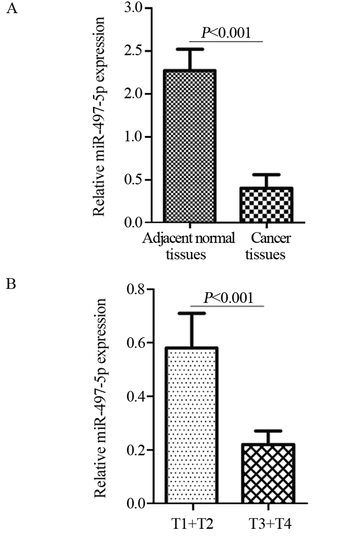
结果miR-497-5p在癌组织中的表达显著低于癌旁正常组织(P < 0.001),T3+T4期患者癌组织中miR-497-5p的表达显著低于T1+T2期癌组织(P < 0.001);低表达miR-497-5p与较高的T分期相关(P=0.003);低表达miR-497-5p的患者5年总体生存率显著低于高表达者(P=0.036)。与对照组相比,miR-497-5p过表达组细胞增殖显著降低,G0/G1期比例增加,S期比例减少。CCNE1 mRNA在癌组织的表达显著高于癌旁正常组织(P < 0.001),且与miR-497-5p表达呈负相关(P < 0.001)。miR-497-5p可直接与CCNE1 mRNA 3’-UTR互补结合从而抑制CCNE1蛋白的表达。
结论miR-497-5p在PaCa中低表达,且与较高的T分期及不良预后相关;过表达miR-497-5p通过靶向调控CCNE1基因诱导细胞周期阻滞进而抑制PaCa细胞增殖。
-
关键词:
- miR-497-5p /
- 胰腺癌 /
- 预后 /
- 增殖 /
- CCNE1
Abstract:ObjectiveTo determine miR-497-5p expression in pancreatic cancer (PaCa) and its clinical significance, and to explore its effects and mechanisms on the proliferation of PaCa cells.
MethodsThe expression levels of miR-497-5p were detected by real-time quantitative PCR experiment. The relation between miR-497-5p expression and clinicopathological features, the prognosis of PaCa patients were analyzed by Chi-square test and Kaplan-Meier method. The effects of miR-497-5p overexpression on the proliferation and cell cycle of Capan-2 and PANC-1 cells were detected by cell counting kit-8 (CCK-8) experiment and flow cytometry. The relation between miR-497-5p level and the cyclin E1 (CCNE1) mRNA expression was analyzed by Spearman correlation test. The regulatory effect of miR-497-5p on CCNE1 expression was confirmed by dual luciferase reporter experiment and Western blot.
ResultsmiR-497-5p expression in cancer tissues was significantly lower than that in adjacent normal tissues (P < 0.001), and its expression in T3+T4 stages cancer tissues were significantly lower than those in T1+T2 stages cancer tissues (P < 0.001); low expression of miR-497-5p was associated with advanced T stage (P=0.003); The 5-year overall survival rate of patients with low miR-497-5p expression was significantly lower than those with high miR-497-5p expression (P=0.036). Compared with the control group, miR-497-5p overexpression significantly reduced cell proliferation, increased the cell proportion in G0/G1 phase and decreased the cell proportion in S phase. The expression levels of CCNE1 mRNA in cancer tissues were significantly higher than those in adjacent normal tissues (P < 0.001), and its expression was negatively correlated with miR-497-5p expression (P < 0.001). miR-497-5p could bind directly to the 3'-UTR of CCNE1 mRNA and inhibit the expression of CCNE1 protein.
ConclusionmiR-497-5p expression is downregulated in PaCa cells and associated with relatively advanced T stage and poor prognosis; the overexpression of miR-497-5p induces the cell cycle arrest to inhibit the proliferation of PaCa cells by targeting CCNE1 gene.
-
Key words:
- miR-497-5p /
- PaCa /
- Prognosis /
- Proliferation /
- CCNE1
-
作者贡献姜达伟:标本收集,实验操作,统计分析,论文撰写与修改许浏:标本收集,统计分析王伟林:实验设计,论文撰写与修改
-
![]()
图 1 实时荧光定量PCR实验分析miR-497-5p在癌与癌旁正常组织(A)及不同T分期癌组织(B)中的表达
Figure 1 miR-497-5p expression in cancer tissues, adjacent normal tissues(A) and different T stages cancer tissues(B) detected by real-time quantitative PCR
![]()
图 2 Kaplan-Meier法分析miR-497-5p的表达与PaCa患者预后的关系
Figure 2 Relation between miR-497-5p expression and prognosis of PaCa patients detected by Kaplan-Meier method
![]()
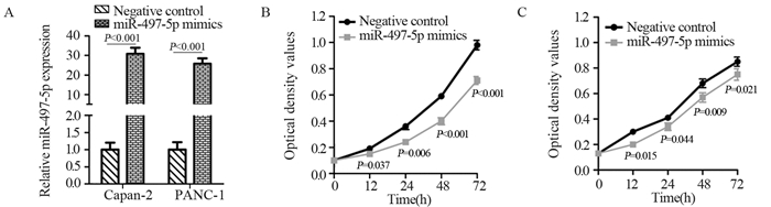
图 3 miR-497-5p模拟物促进miR-497-5p的表达(A)并抑制Capan-2(B)和PANC-1(C)细胞的增殖
Figure 3 miR-497-5p mimics promoted miR-497-5p expression(A) and inhibited proliferation of Capan-2(B) and PANC-1(C) cells
![]()
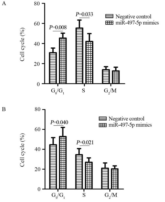
图 4 miR-497-5p模拟物对Capan-2(A)和PANC-1(B)细胞周期的影响
Figure 4 Effect of miR-497-5p mimics on cell cycle of Capan-2(A) and PANC-1(B) cells
![]()
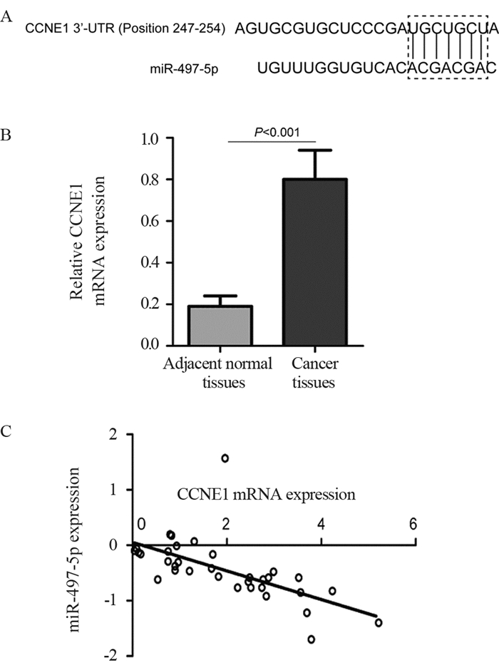
图 5 生物信息学分析miR-497-5p对CCNE1的调控作用
Figure 5 Effect of miR-497-5P on CCNE1 bioinformatics analysis
![]()
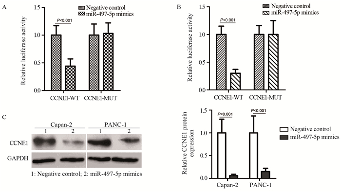
图 6 miR-497-5p对CCNE1基因的调控作用分析
Figure 6 Analysis of the regulatory effect of miR-497-5p on CCNE1 gene
表 1 miR-497-5p的表达与胰腺癌患者临床病理特征之间的关系
Table 1 Relation between miR-497-5p expression and clinicopathological characteristics of pancreatic cancer (PaCa) patients

 下载: 导出CSV
-
[1] Siegel RL, Miller KD, Jemal A. Cancer Statistics, 2017[J]. CA Cancer J Clin, 2017, 67(1): 7-30. doi: 10.3322/caac.21387
[2] Ilic M, Ilic I. Epidemiology of pancreatic cancer[J]. World J Gastroenterol, 2016, 22(44): 9694-9705. doi: 10.3748/wjg.v22.i44.9694
[3] Yonemori K, Kurahara H, Maemura K, et al. MicroRNA in pancreatic cancer[J]. J Hum Genet, 2017, 62(1): 33-40. http://www.ncbi.nlm.nih.gov/pubmed/27251005
[4] 何峰, 陈曦, 程方雄, 等. miR-130b调控RAB34的表达在胰腺癌发生发展中的作用[J].肿瘤防治研究, 2018, 45(3): 144-147. doi: 10.3971/j.issn.1000-8578.2018.17.0960 He F, Chen X, Cheng FX, et al. Effect of miR-130b Targeting RAB34 Expression in Occurrence and Development of Pancreatic Cancer[J]. Zhong Liu Fang Zhi Yan Jiu, 2018, 45(3): 144-147. doi: 10.3971/j.issn.1000-8578.2018.17.0960
[5] Chen Y, Kuang D, Zhao X, et al. miR-497-5p inhibits cell proliferation and invasion by targeting KCa3.1 in angiosarcoma[J]. Oncotarget, 2016, 7(36): 58148-58161. doi: 10.18632/oncotarget.11252
[6] Sun Z, Li A, Yu Z, et al. MicroRNA-497-5p Suppresses Tumor Cell Growth of Osteosarcoma by Targeting ADP Ribosylation Factor-Like Protein 2[J]. Cancer Biother Radiopharm, 2017, 32(10): 371-378. doi: 10.1089/cbr.2017.2268
[7] Chai L, Kang XJ, Sun ZZ, et al. MiR-497-5p, miR-195-5p and miR-455-3p function as tumor suppressors by targeting hTERT in melanoma A375 cells[J]. Cancer Manag Res, 2018, 10: 989-1003. doi: 10.2147/CMAR.S163335
[8] Ran X, Xiao CH, Xiang GM, et al. Regulation of Embryonic Stem Cell Self-Renewal and Differentiation by MicroRNAs[J]. Cell Reprogram, 2017, 19(3): 150-158. http://cn.bing.com/academic/profile?id=21aa027f7158bd8d4d66a6c5f000160e&encoded=0&v=paper_preview&mkt=zh-cn
[9] Quinlan S, Kenny A, Medina M, et al. MicroRNAs in Neurodegenerative Diseases[J]. Int Rev Cell Mol Biol, 2017, 334: 309-343. doi: 10.1016/bs.ircmb.2017.04.002
[10] Nemecz M, Alexandru N, Tanko G, et al. Role of MicroRNA in Endothelial Dysfunction and Hypertension[J]. Curr Hypertens Rep, 2016, 18(12): 87. doi: 10.1007/s11906-016-0696-8
[11] Agrawal S. Potential prognostic biomarkers in pancreatic juice of resectable pancreatic ductal adenocarcinoma[J]. World J Clin Oncol, 2017, 8(3): 255-260. doi: 10.5306/wjco.v8.i3.255
[12] Svoronos AA, Engelman DM, and Slack FJ. OncomiR or Tumor Suppressor? The Duplicity of MicroRNAs in Cancer[J]. Cancer Res, 2016, 76(13): 3666-3670. doi: 10.1158/0008-5472.CAN-16-0359
[13] Lv F, Zheng K, Yu J, et al. MicroRNA-661 expression is upregulated in pancreatic ductal adenocarcinoma and promotes cell proliferation[J]. Oncol Lett, 2018, 16(5): 6293-6298. http://cn.bing.com/academic/profile?id=2f71508910f067709c9e54b41c89f0e6&encoded=0&v=paper_preview&mkt=zh-cn
[14] Ta N, Huang X, Zheng K, et al. miRNA-1290 Promotes Aggressiveness in Pancreatic Ductal Adenocarcinoma by Targeting IKK1[J]. Cell Physiol Biochem, 2018, 51(2): 711-728. http://cn.bing.com/academic/profile?id=0096288dd8183f53cd14be6d89c3ae27&encoded=0&v=paper_preview&mkt=zh-cn
[15] Zhu L, Chen Y, Nie K, et al. MiR-101 inhibits cell proliferation and invasion of pancreatic cancer through targeting STMN1[J]. Cancer Biomark, 2018, 23(2): 301-309. doi: 10.3233/CBM-181675
[16] Yang Y, Tao X, Li CB, et al. MicroRNA-494 acts as a tumor suppressor in pancreatic cancer, inhibiting epithelial-mesenchymal transition, migration and invasion by binding to SDC1[J]. Int J Oncol, 2018, 53(3): 1204-1214. http://cn.bing.com/academic/profile?id=f1549a5f52fc6d3f041c065019a73a7a&encoded=0&v=paper_preview&mkt=zh-cn
[17] Li G, Wang K, Wang J, et al. miR-497-5p inhibits tumor cell growth and invasion by targeting SOX5 in non-small-cell lung cancer[J]. J Cell Biochem, 2019, 120(6): 10587-10595. doi: 10.1002/jcb.28345
[18] Zhao H, Wang J, Zhang Y, et al. Prognostic Values of CCNE1 Amplification and Overexpression in Cancer Patients: A Systematic Review and Meta-analysis[J]. J Cancer, 2018, 9(13): 2397-2407. doi: 10.7150/jca.24179
[19] de Maturana EL, Rava M, Anumudu C, et al. Bladder Cancer Genetic Susceptibility. A Systematic Review[J]. Bladder Cancer, 2018, 4(2): 215-226. doi: 10.3233/BLC-170159

计量
- 文章访问数: 2262
- HTML全文浏览量: 516
- PDF下载量: 519

