
SerpinA5 Inhibits Malignant Biological Behavior of Esophageal Squamous Cell Carcinoma by Regulating Fn/Integrin-β1 Signaling Pathway
-
摘要:目的
探讨SerpinA5对食管鳞癌(ESCC)细胞恶性生物学行为的影响及其分子机制。
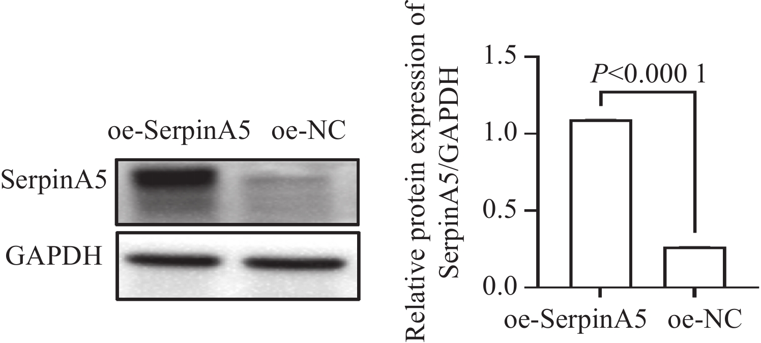
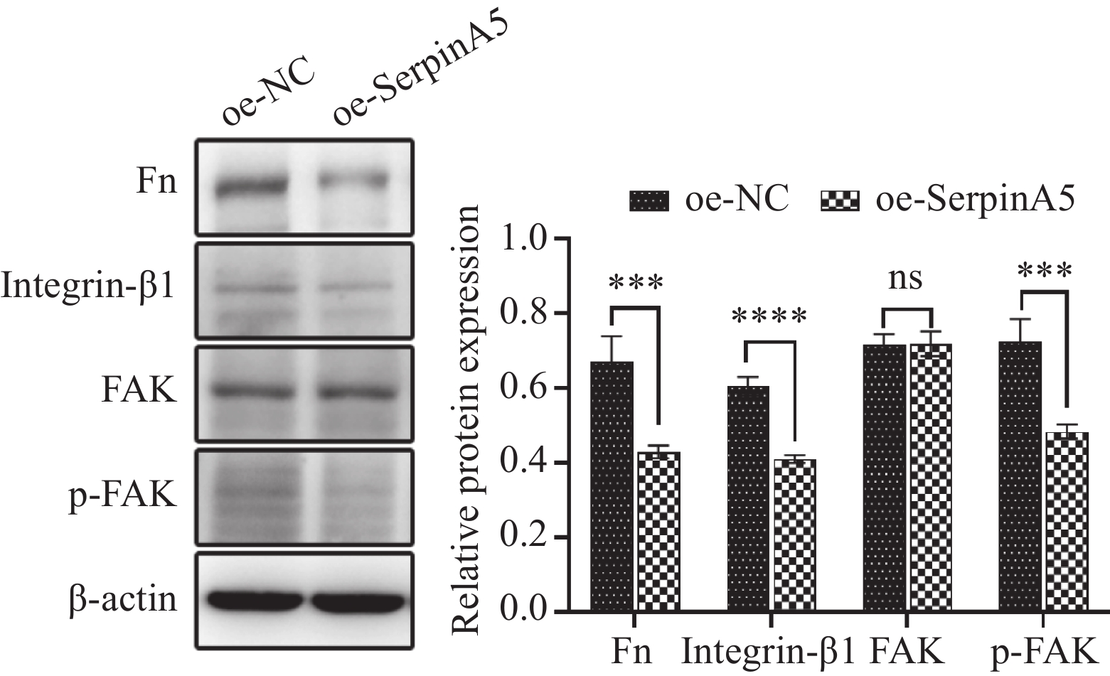
方法通过TIMER2.0数据库分析SerpinA5基因在不同肿瘤和相邻正常组织之间的表达水平。Western blot检测SerpinA5在ESCC细胞系和食管上皮细胞中的表达情况。利用慢病毒构建SerpinA5过表达KYSE150细胞稳转株,Western blot方法检测过表达效率。采用CCK8、平板克隆实验、流式细胞术、创面愈合实验、Transwell侵袭实验检测过表达SerpinA5对食管鳞癌细胞增殖、凋亡、迁移及侵袭能力的影响。构建过表达SerpinA5的裸鼠皮下移植瘤模型。观察肿瘤生长,测量瘤体的体积和质量。IHC法检测裸鼠皮下移植瘤中细胞增殖水平。采用免疫共沉淀(Co-IP)方法明确SerpinA5与Fn之间的相互作用。Western blot方法检测移植瘤中Fn/Integrin-β1信号通路相关蛋白(Fn、Integrin-β1、FAK和p-FAK)的表达水平变化。
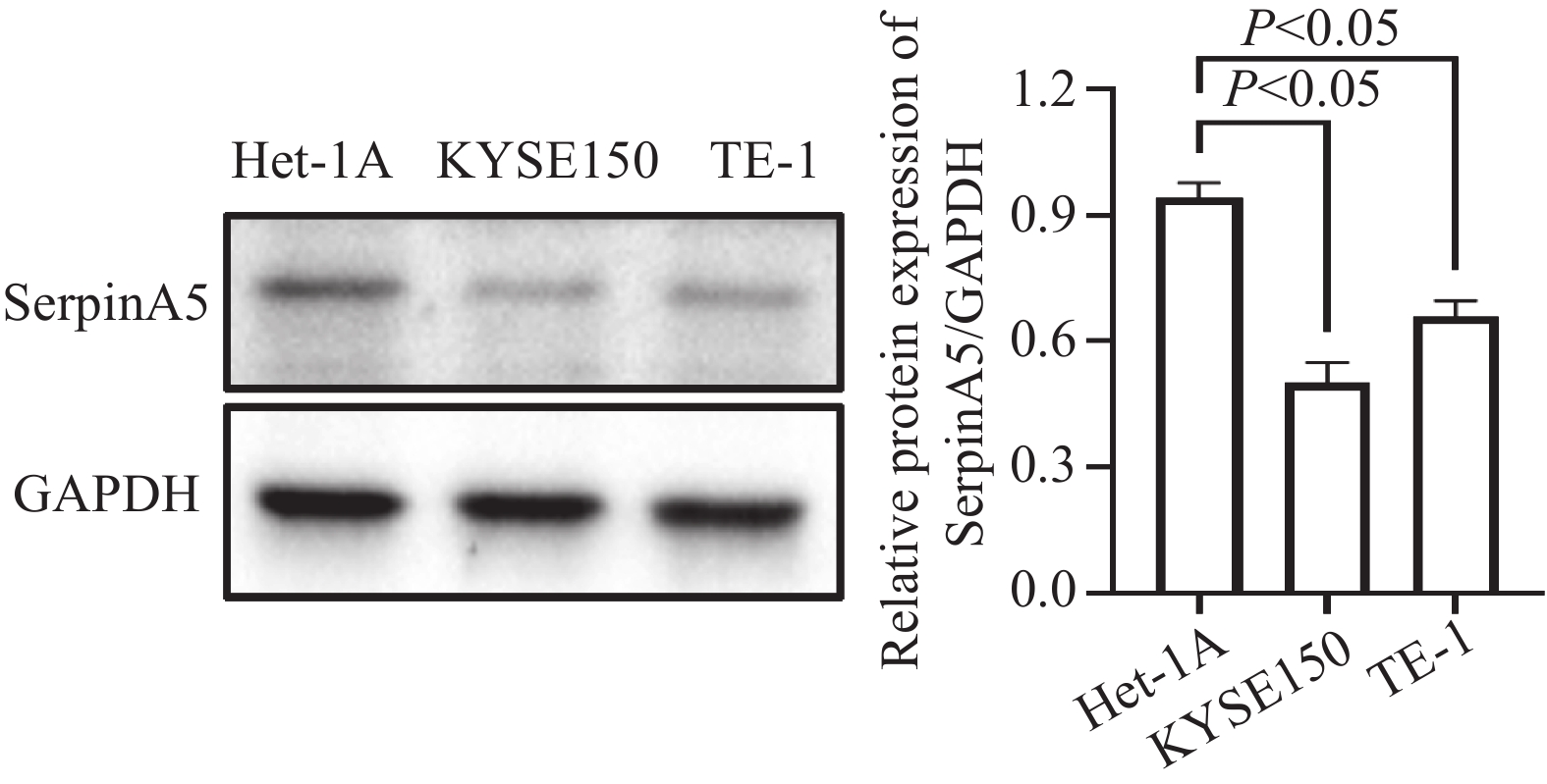
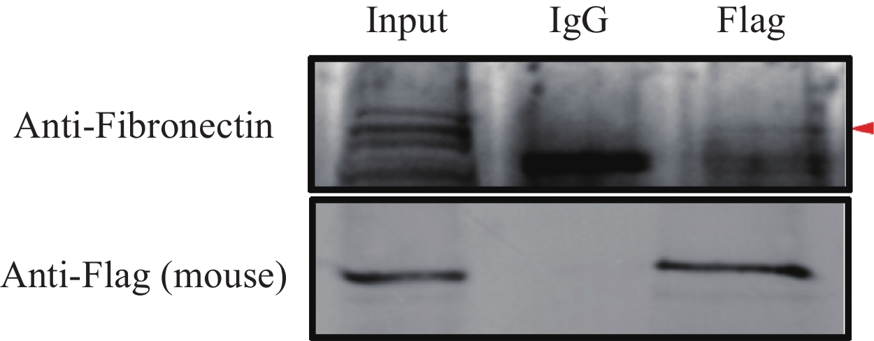
结果SerpinA5在ESCC组织及细胞系中均为低表达水平。在ESCC细胞中过表达SerpinA5后,可显著抑制细胞的增殖、迁移及侵袭,促进其凋亡。SerpinA5过表达组的瘤体体积和质量均小于阴性对照组。IHC结果显示SerpinA5过表达可显著抑制瘤体中ESCC细胞增殖。Co-IP证实SerpinA5与Fn存在相互作用。过表达SerpinA5后裸鼠皮下移植瘤内ESCC细胞中Fn/Integrin-β1信号通路相关蛋白Fn、Integrin-β1、p-FAK表达水平显著降低。
结论Serpin A5可能通过调控Fn/Integrin-β1信号通路抑制食管鳞癌细胞的增殖、迁移、侵袭,并促进其凋亡。
-
关键词:
- 食管鳞状细胞癌 /
- SerpinA5 /
- 恶性生物学行为 /
- Fn/Integrin-β1信号通路
Abstract:ObjectiveTo investigate the effect of SerpinA5 on the malignant biological behavior of esophageal squamous cell carcinoma (ESCC) and its molecular mechanism.
MethodsThe expression levels of the SerpinA5 gene in various tumors and adjacent normal tissues were analyzed by using the TIMER2.0 database. The expression levels of SerpinA5 in the ESCC cell line and esophageal epithelial cells were detected through Western blot analysis. Stably transfected KYSE150 cell line with overexpression of SerpinA5 was constructed through lentiviral transfection, and overexpression efficiency was detected via Western blot analysis. The effects of SerpinA5 overexpression on the proliferation, apoptosis, migration, and invasion of ESCC cells were detected by employing the CCK8, plate cloning, flow cytometry, wound healing, and Transwell invasion assays. The nude mice subcutaneous xenograft model with SerpinA5 overexpression was constructed. Tumor growth was observed, and tumor volume and mass were measured. The cell proliferation level of the subcutaneous xenograft tumors in nude mice was detected via immunohistochemistry (IHC). Coimmunoprecipitation (Co-IP) was employed to determine the interaction between SerpinA5 and Fn. Western blot analysis was applied to detect the expression levels of proteins (Fn, Integrin-β1, FAK, and p-FAK) related to the Fn/Integrin-β1 signaling pathway in transplanted tumors.
ResultsSerpinA5 was expressed at low levels in ESCC tissues and cell lines. In ESCC cells, SerpinA5 overexpression can considerably inhibit cell proliferation, migration, and invasion and promote cell apoptosis. In the subcutaneous xenograft experiment on nude mice, the tumor volume and weight of the SerpinA5 overexpression group were lower than those of the negative control group. IHC results demonstrated that SerpinA5 overexpression significantly inhibited the proliferation of ESCC cells in tumor tissues. Co-IP confirmed the interaction between SerpinA5 and Fn. Western blot analysis results showed that the expression levels of Fn, Integrin-β1, and p-FAK in the Fn/Integrin-β1 signaling pathway of ESCC cells in the subcutaneous xenograft tumors of nude mice significantly decreased after SerpinA5 overexpression.
ConclusionSerpin A5 may inhibit proliferation, migration, and invasion and promote apoptosis of ESCC cells by regulating the Fn/Integrin-β1 signaling pathway.
-
Competing interests: The authors declare that they have no competing interests.利益冲突声明:所有作者均声明不存在利益冲突。作者贡献:魏 瑜:动物实验、数据分析及论文撰写张周华、李志芳:生信分析及细胞实验张 莉:实验设计及论文审校
-
![]()
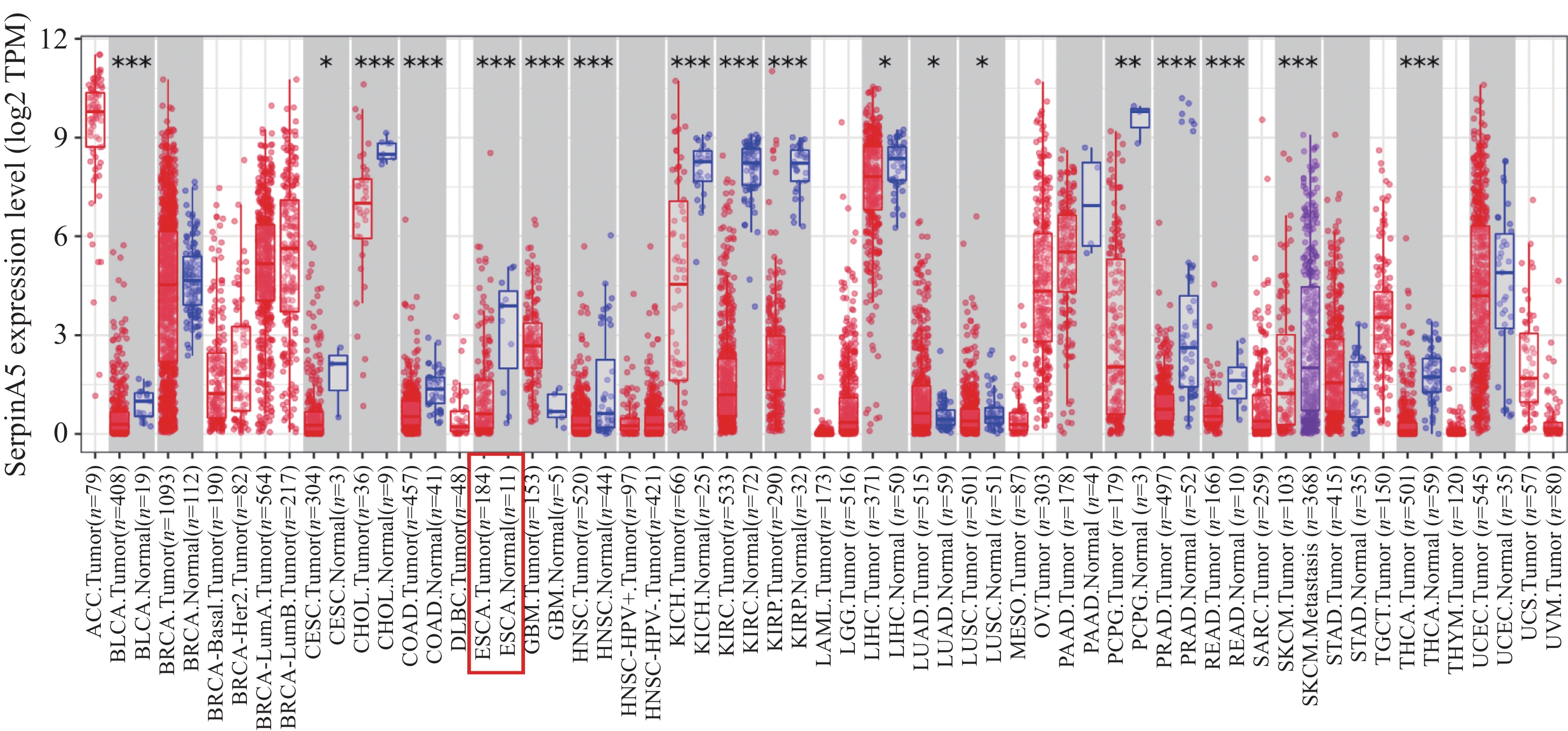
图 1 TIMER2.0数据库中SerpinA5 mRNA在不同肿瘤中的表达情况
Figure 1 Expression levels of SerpinA5 mRNA in different tumors in the TIMER2.0 database
![]()
图 2 Western blot检测SerpinA5在食管鳞癌细胞及食管上皮细胞中的表达
Figure 2 Expression levels of SerpinA5 in ESCC and esophageal epithelial cell lines detected by Western blot analysis
![]()
图 3 Western blot检测食管鳞癌细胞SerpinA5过表达慢病毒的转染效率
Figure 3 Transfection efficiency of lentiviral vectors for SerpinA5 overexpression in ESCC cells detected by Western blot analysis
![]()
图 4 SerpinA5基因过表达对KYSE150细胞增殖(A、B)、凋亡(C)、迁移(D)、侵袭(E)能力的影响
Figure 4 Effects of SerpinA5 overexpression on the proliferation (A, B), apoptosis (C), migration (D), and invasion (E) of KYSE150 cells
![]()
图 7 Co-IP法验证在食管鳞癌细胞中SerpinA5与Fn相互作用
Figure 7 Verification of the interaction between SerpinA5 and Fn in ESCC cells by Co-IP
-
[1] Ferlay J, Ervik M, Lam F, et al. Globocan 2022 (version 1.1) [EB/OL]. https://gco.iarc.who.int/today/en/dataviz/bars?mode=cancer&group_populations=1&populations=900&types=1&sort_by=value0&key=total. –[2024-02-08].
[2] Xu QL, Li H, Zhu YJ, et al. The treatments and postoperative complications of esophageal cancer: a review[J]. J Cardiothorac Surg, 2020, 15(1): 163-173. doi: 10.1186/s13019-020-01202-2
[3] 郑荣寿, 陈茹, 韩冰峰, 等. 2022年中国恶性肿瘤流行情况分析[J]. 中华肿瘤杂志, 2024, 46(3): 221-231. [Zheng RS, Chen R, Han BF, et al. Cancer incidence and mortality in China, 2022[J]. Zhonghua Zhong Liu Za Zhi, 2024, 46(3): 221-231.] doi: 10.3760/cma.j.cn112152-20240119-00035 Zheng RS, Chen R, Han BF, et al. Cancer incidence and mortality in China, 2022[J]. Zhonghua Zhong Liu Za Zhi, 2024, 46(3): 221-231. doi: 10.3760/cma.j.cn112152-20240119-00035
[4] Thrift AP. The epidemic of oesophageal carcinoma: Where are we now?[J]. Cancer Epidemiol, 2016, 41: 88-95. doi: 10.1016/j.canep.2016.01.013
[5] Zeng H, Chen W, Zheng R, et al. Changing cancer survival in China during 2003-15: a pooled analysis of 17 population-based cancer registries[J]. Lancet Glob Health, 2018, 6(5): e555-e567. doi: 10.1016/S2214-109X(18)30127-X
[6] 陈星. SERPINA5预测食管鳞癌放化疗疗效及预后的价值[D]. 乌鲁木齐:新疆医科大学, 2020. [Chen X. The value of SERPINA5 in predicting the efficacy and prognosis of radiotherapy and chemotherapy in esophageal squamous cell carcinoma[D]. Urumqi: Xinjiang Medical University, 2020.] Chen X. The value of SERPINA5 in predicting the efficacy and prognosis of radiotherapy and chemotherapy in esophageal squamous cell carcinoma[D]. Urumqi: Xinjiang Medical University, 2020.
[7] Yang H, Wahlmüller FC, Sarg B, et al. A+-helix of protein C inhibitor (PCI) is a cell-penetrating peptide that mediates cell membrane permeation of PCI[J]. J Biol Chem, 2015, 290(5): 3081-3091. doi: 10.1074/jbc.M114.581736
[8] Wahlmüller FC, Yang H, Furtmüller M, et al. Regulation of the Extracellular SERPINA5(Protein C Inhibitor) Penetration Through Cellular Membranes[J]. Adv Exp Med Biol, 2017, 966: 93-101.
[9] Wakita T, Hayashi T, Nishioka J, et al. Regulation of carcinoma cell invasion by protein C inhibitor whose expression is decreased in renal cell carcinoma[J]. Int J Cancer, 2004, 108(4): 516-523. doi: 10.1002/ijc.11594
[10] Song Y, Ye L, Tan Y, et al. Therapeutic exosomes loaded with SERPINA5 attenuated endometrial cancer cell migration via the integrin β1/FAK signaling pathway[J]. Cell Oncol (Dordr), 2022, 45(5): 861-872.
[11] Jing Y, Jia D, Wong CM, et al. SERPINA5 inhibits tumor cell migration by modulating the fibronectin-integrin β1 signaling pathway in hepatocellular carcinoma[J]. Mol Oncol, 2014, 8(2): 366-377. doi: 10.1016/j.molonc.2013.12.003
[12] Morgan E, Soerjomataram I, Gavin A T, et al. International trends in oesophageal cancer survival by histological subtype between 1995 and 2014[J]. Gut, 2021, 70(2): 234-242.
[13] Asanuma K, Yoshikawa T, Hayashi T, et al. Protein C inhibitor inhibits breast cancer cell growth, metastasis and angiogenesis independently of its protease inhibitory activity[J]. Int J Cancer, 2007, 121(5): 955-965. doi: 10.1002/ijc.22773
[14] Bijsmans Ingrid TGW, Smits Kim M, de Graeff Pauline, et al. Loss of SerpinA5 protein expression is associated with advanced-stage serous ovarian tumors[J]. Mod Pathol, 2011, 24: 463-470.
[15] Palmieri D, Lee JW, Juliano RL, et al. Plasminogen activator inhibitor-1 and -3 increase cell adhesion and motility of mda-mb-435 breast cancer cells[J]. J Biol Chem, 2002, 277(43): 40950-40957. doi: 10.1074/jbc.M202333200
[16] Fan M, Xiong X, Han L, et al. Serpina5 promotes tumour cell proliferation by modulating the pi3k/akt/mtor signalling pathway in gastric cancer[J]. J Cell Mol Med, 2022, 26(18): 4837-4846. doi: 10.1111/jcmm.17514
[17] Zhang L, Hu S, Korteweg C, et al. Expression of immunoglobulin G in esophageal squamous cell carcinomas and its association with tumor grade and Ki67[J]. Hum Pathol, 2012, 43(3): 423-434. doi: 10.1016/j.humpath.2011.05.020
[18] Ma H, Wang J, Zhao X, et al. Periostin Promotes Colorectal Tumorigenesis through Integrin-FAK-Src Pathway-Mediated YAP/TAZ Activation[J]. Cell Rep, 2020, 30(3): 793-806. e6.
[19] Kuonen F, Surbeck I, Sarin KY, et al. TGFβ, Fibronectin and Integrin α5β1 Promote Invasion in Basal Cell Carcinoma[J]. J Invest Dermatol, 2018, 138(11): 2432-2442. doi: 10.1016/j.jid.2018.04.029
[20] Vega ME, Schwarzbauer JE. Collaboration of Fibronectin Matrix with other Extracellular Signals in Morphogenesis and Differentiation[J]. Curr Opin Cell Biol, 2016, 42: 1-6. doi: 10.1016/j.ceb.2016.03.014
[21] Strohmeyer N, Bharadwaj M, Costell M, et al. Fibronectin-bound α5β1 integrins sense load and signal to reinforce adhesion in less than a second[J]. Nat Mater, 2017, 16(12): 1261-1270.
[22] Li K, Zhao G, Ao J, et al. ZNF32 induces anoikis resistance through maintaining redox homeostasis and activating Src/FAK signaling in hepatocellular carcinoma[J]. Cancer Lett, 2019, 442: 271-278. doi: 10.1016/j.canlet.2018.09.033
[23] Zhao G, Gong L, Su D, et al. Cullin5 deficiency promotes small-cell lung cancer metastasis by stabilizing integrin β1[J]. J Clin Invest, 2019, 129(3): 971-987.
 下载:
下载:


计量
- 文章访问数: 2002
- HTML全文浏览量: 3208
- PDF下载量: 186

