
头颈部脂肪肉瘤预后相关因素分析
作者简介:
丁硕,女,博士,主治医师,主要从事头颈部肿瘤常规及微创治疗研究,ORCID: 0000-0002-9600-043X
 钟琦: 博士,主任医师,教授,硕士生导师,长期从事头颈部肿瘤的常规、微创治疗及头颈部缺损的手术修复重建工作。中国残疾人康复协会无喉者康复专业委员会主任委员;中国医疗保健国际交流促进会鼻咽癌防治分会副秘书长、委员;中国医疗保健国际交流促进会甲状腺疾病防治分会副主任委员、副秘书长;中华志愿者协会中西医结合专家志愿者委员会耳鼻喉头颈外科专业组副组长;北京癌症防治学会头颈肿瘤MDT分会副主任委员;北京市医学会耳鼻咽喉头颈外科专业委员会秘书;《中国医药科学》杂志编委;《中华耳鼻咽喉头颈外科杂志》审稿人、通讯编委;《中国肿瘤整合诊治指南(CACA):头颈肿瘤(2022)》副主编;《头颈诊断病理学》主译。2017年获得残疾预防及康复科学技术奖三等奖;2020年获得华夏医疗保健国际交流促进科技奖二等奖;2021年获得“亦麒麟”优秀人才;2023年获得中华医学科学技术奖二等奖。主持10项市级及国家级课题,参与国家级课题10余项目,发表中文核心期刊论文30余篇,发表SCI论文20余篇
。
钟琦: 博士,主任医师,教授,硕士生导师,长期从事头颈部肿瘤的常规、微创治疗及头颈部缺损的手术修复重建工作。中国残疾人康复协会无喉者康复专业委员会主任委员;中国医疗保健国际交流促进会鼻咽癌防治分会副秘书长、委员;中国医疗保健国际交流促进会甲状腺疾病防治分会副主任委员、副秘书长;中华志愿者协会中西医结合专家志愿者委员会耳鼻喉头颈外科专业组副组长;北京癌症防治学会头颈肿瘤MDT分会副主任委员;北京市医学会耳鼻咽喉头颈外科专业委员会秘书;《中国医药科学》杂志编委;《中华耳鼻咽喉头颈外科杂志》审稿人、通讯编委;《中国肿瘤整合诊治指南(CACA):头颈肿瘤(2022)》副主编;《头颈诊断病理学》主译。2017年获得残疾预防及康复科学技术奖三等奖;2020年获得华夏医疗保健国际交流促进科技奖二等奖;2021年获得“亦麒麟”优秀人才;2023年获得中华医学科学技术奖二等奖。主持10项市级及国家级课题,参与国家级课题10余项目,发表中文核心期刊论文30余篇,发表SCI论文20余篇
。
通信作者:
-
摘要:目的
探讨头颈部区域脂肪肉瘤的发病以及预后影响因素,同时分析不同治疗方案的疗效。
方法回顾性分析2008年1月至2024年1月本院接治的头颈部脂肪肉瘤患者。所有患者均接受随访,利用SPSS软件分析患者预后。
结果共纳入患者30例,其中脂肪肉瘤在眼眶内可达60%,其余脂肪肉瘤主要分布在颈深部各个间隙中。去分化脂肪肉瘤(33%)最常见,黏液多形性脂肪肉瘤(4%)最少见。肿瘤病理类型(P<0.001)和Ki67(P=0.014)对肿瘤控制率有显著影响。针对肿瘤的疾病特异性生存率进行分析,显示各项因素间差异无统计学意义(均P>0.05)。
结论头颈部脂肪肉瘤的预后相对于其他部位的脂肪肉瘤预后更好,但黏液多形性脂肪肉瘤或多形性脂肪肉瘤以及Ki67过高是预后差的信号,而术后的辅助放射治疗并不能显著提高疾病特异性生存率。
Abstract:ObjectiveTo explore the pathogenesis and prognostic factors of liposarcoma in the head and neck region, and simultaneously analyze the efficacy of different treatment regimens.
MethodsA retrospective analysis was performed on all patients with primary untreated head and neck liposarcoma who were diagnosed and underwent surgical treatment at our hospital from January 2008 to January 2024. All patients were monitored during follow-up, and their prognoses were analyzed using SPSS software.
ResultsA total of 30 patients were included in the study. Liposarcoma accounted for up to 60% of the cases in the orbit, while the remaining liposarcomas were primarily located in various interspaces of the neck. Dedifferentiated liposarcoma was the most common type, comprising 33%, while myxoid pleomorphic liposarcoma was the rarest at 4%. The tumor pathological type (P<0.001) and Ki67 (P=0.014) significantly affected the tumor control rate. However, an analysis of disease-specific survival rates revealed no significant differences across various factors (all P>0.05).
ConclusionThe prognosis of head and neck liposarcoma is better compared to that of liposarcomas in other parts of the body. However, myxoid pleomorphic liposarcoma, pleomorphic fat sarcoma, and high Ki67 levels are indicators of poor prognosis. Additionally, postoperative adjuvant radiotherapy does not significantly enhance disease-specific survival rates.
-
Key words:
- Head and neck /
- Liposarcoma /
- Prognosis analysis /
- Treatment
-
Competing interests: The authors declare that they have no competing interests.利益冲突声明:所有作者均声明不存在利益冲突。作者贡献:丁 硕:文章撰写与修改黄志刚、张洋:提出研究选题、设计论文框架侯丽珍、郭伟:整理数据尹高菲:调研整理文献房居高、钟琦:文章审阅
-
![]()
图 1 Ki67表达水平对脂肪肉瘤患者局部控制率的影响
Figure 1 Influence of Ki67 expression level on local control rate of liposarcoma patients
![]()
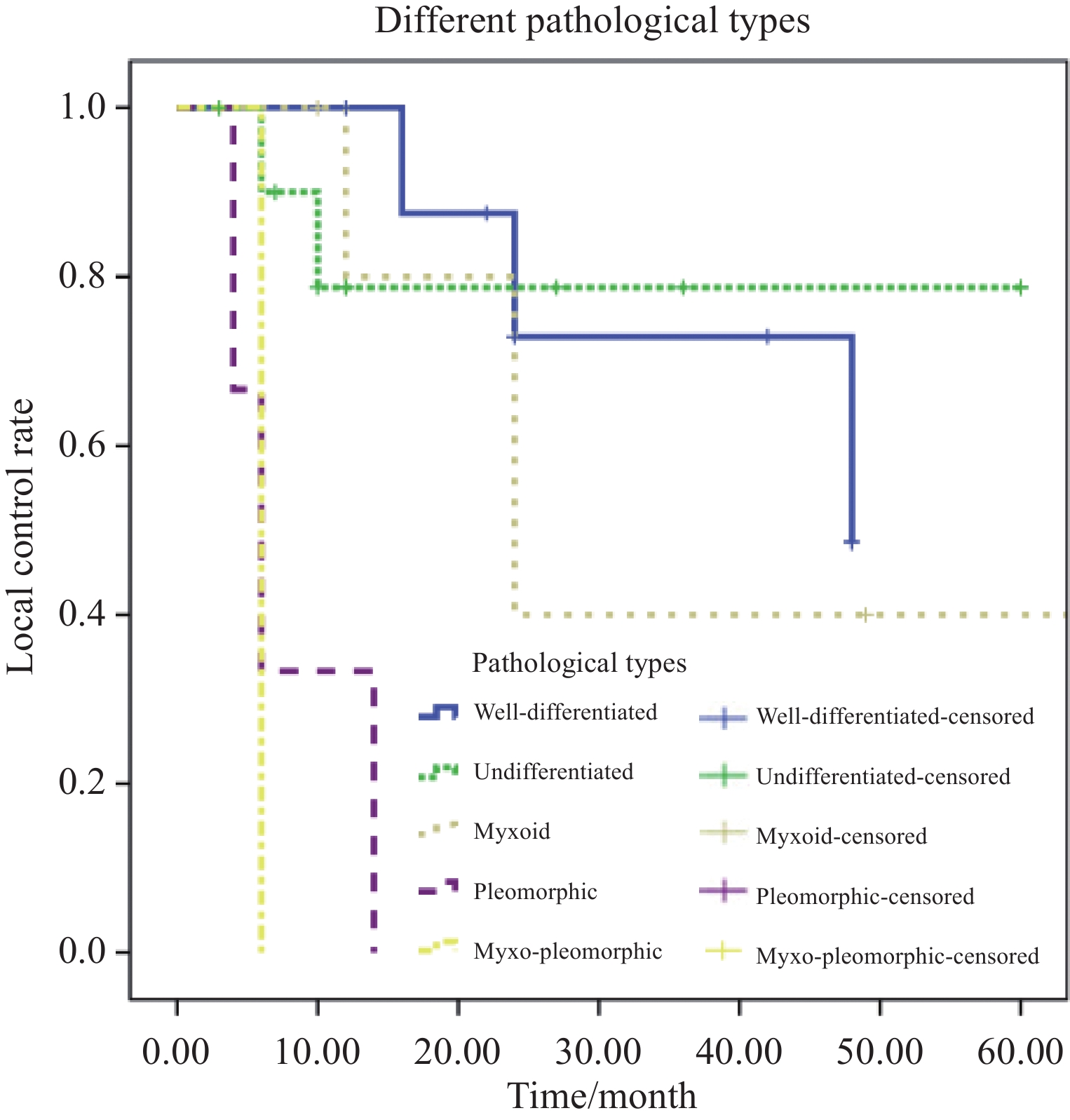
图 2 病理类型对脂肪肉瘤患者局部控制率的影响
Figure 2 Effect of pathological subtype on local control rate of liposarcoma patients
![]()
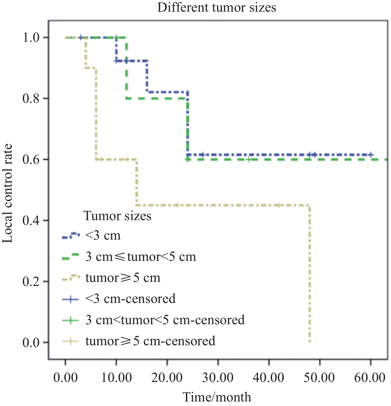
图 3 肿瘤大小对脂肪肉瘤患者局部控制率的影响
Figure 3 Effect of tumor size on local control rate of liposarcoma patients
表 1 脂肪肉瘤发病部位及病理类型
Table 1 Location and pathological type of liposarcoma
Site Pathological type Total Well-differentiated Undifferentiated Myxoid Pleomorphic Myxo-pleomorphic Orbital cavity 4 7 6 1 0 18 Retropharyngeal space 2 1 0 0 0 3 Carotid space 1 1 0 2 0 4 Parapharyngeal space 0 1 0 0 0 1 Submental space 1 0 0 0 0 1 Larynx and hypopharynx 1 1 0 0 0 2 Temporal fossa 0 0 0 0 1 1 Total 9 11 6 3 1 30  下载: 导出CSV
表 2 脂肪肉瘤的治疗及病理类型
Table 2 Treatment and pathologic types of liposarcoma
Treatment Pathological type Total Well-
differentiatedUndiffer-
entiatedMyxoid Pleom-
orphicMyxo-
pleomorphicSurgery 6 7 0 3 0 16 Surgery+
radiation2 3 4 1 1 11 Surgery+
particle
therapy1 0 2 0 0 3 Total 9 10 6 4 1 30
下载: 导出CSV
表 3 接受不同治疗方式的脂肪肉瘤患者随访结果
Table 3 Follow-up of patients with liposarcoma treated with different modalities
Treatment No. Survival Recurrence Metastasis Surgery 16 15 6 0 Surgery+radiation 11 8 6 1 Surgery+particle 3 3 0 0 Total 30 2 12 1
下载: 导出CSV
表 4 脂肪肉瘤患者临床病理特征与预后的相关性
Table 4 Correlation between clinical pathological features and prognosis in patients with liposarcoma
Pathological
featuresn Percentage
(%)Control
rateP Disease-free
survivalP Gender 0.532 0.314 Male 12 40 38.1 60.6 Female 18 60 54.9 83.7 Site 0.651 0.940 Orbital set 18 60 45.6 77.2 Nonorbital
set12 40 47.6 68.8 Age (years) 0.208 0.073 ≥18-45 13 43 27.8 90.0 ≥45-60 10 33 42.9 68.6 ≥60 7 24 75.0 62.5 History of
benign
mesenchymal
origin tumors0.153 0.276 Yes 8 27 18.8 100.0 No 22 73 55.8 83.0 Tumor
size(cm)0.065 0.851 <3 14 47 61.0 92.0 3-5 6 20 60.0 75.0 ≥5 10 33 10.0 90.0 Ki67 0.014 0.169 <10% 16 54 56.4 100.0 10%-30% 7 23 56.6 87.5 ≥30%-50% 3 10 0.0 50.0 ≥50% 4 13 0.0 66.7 Surgery option 0.311 0.349 Extended resection 5 17 49.6 80.0 Tumor excision 25 83 30.0 89.7 Tumor capsule 0.945 0.12 Yes 17 57 50.3 100.0 No 13 43 46.3 78.8 Postoperative treatment 0.116 0.087 Yes 14 47 57.6 77.1 No 16 53 41.6 100.0 Pathological type <0.001 0.187 Well- differentiated 9 30 48.6 100.0 Dedifferentiated liposarcoma 10 33 68.8 100.0 Myxoid 6 20 40.0 66.7 Pleomorphic 4 13 0.0 66.7 Myxo- pleomorphic 1 4 0.0 0.0
下载: 导出CSV
-
[1] Sbaraglia M, Bellan E, Dei Tos AP. The 2020 WHO Classification of Soft Tissue Tumours: news and perspectives[J]. Pathologica, 2021, 113(2): 70-84.
[2] de Bree R, van der Waal I, de Bree E, et al. Management of adult soft tissue sarcomas of the head and neck[J]. Oral Oncol, 2010, 46(11): 786-790. doi: 10.1016/j.oraloncology.2010.09.001
[3] Ramu EM, Houdek MT, Isaac CE, et al. Management of soft-tissue sarcomas; treatment strategies, staging, and outcomes[J]. SICOT-J, 2017, 3: 20. doi: 10.1051/sicotj/2017010
[4] Mastrangelo G, Coindre JM, Ducimetière F, et al. Incidence of soft tissue sarcoma and beyond: a population-based prospective study in 3 European regions[J]. Cancer, 2012, 118(21): 5339-5348. doi: 10.1002/cncr.27555
[5] Luo P, Cai W, Yang L, et al. Retroperitoneal dedifferentiated liposarcoma: Analysis of 61 cases from a large institution[J]. J Cancer, 2018, 9(21): 3831-3838. doi: 10.7150/jca.25715
[6] Furukawa H, Tanemura M, Matsuda H, et al. A giant esophageal liposarcoma radically resected by the cervical approach: a case report[J]. Clin J Gastroenterol, 2022, 15(1): 71-76. doi: 10.1007/s12328-021-01550-z
[7] Barisella M, Giannini L, Piazza C. From head and neck lipoma to liposarcoma: a wide spectrum of differential diagnoses and their therapeutic implications[J]. Curr Opin Otolaryngol Head Neck Surg, 2020, 28(2): 136-143. doi: 10.1097/MOO.0000000000000608
[8] Lawrence W Jr, Anderson JR, Gehan EA, et al. Pretreatment TNM staging of childhood rhabdomyosarcoma: A report of the Intergroup Rhabdomyosarcoma Study Group. Children's Cancer Study Group. Pediatric Oncology Group[J]. Cancer, 1997, 80(6): 1165-1170. doi: 10.1002/(SICI)1097-0142(19970915)80:6<1165::AID-CNCR21>3.0.CO;2-5
[9] Choi JH, Ro JY. The 2020 WHO Classification of Tumors of Soft Tissue: Selected Changes and New Entities[J]. Adv Anat Pathol, 2021, 28(1): 44-58. doi: 10.1097/PAP.0000000000000284
[10] Yang T, Xiao L, Ren H. A well-differentiated liposarcoma of the prevertebral space: a case report[J]. Transl Cancer Res, 2021, 10(7): 3600-3604. doi: 10.21037/tcr-21-143
[11] Gerry D, Fox NF, Spruill LS, et al. Liposarcoma of the head and neck: Analysis of 318 cases with comparison to non–head and neck sites[J]. Head Neck, 2014, 36(3): 393-400. doi: 10.1002/hed.23311
[12] Nili F, Baghai F, Aghai A, et al. Well-differentiated liposarcoma of the floor of the mouth: Report of a rare case and review of the literature[J]. J Oral Maxillofac Pathol, 2016, 20(2): 312-315. doi: 10.4103/0973-029X.185984
[13] Mouret P. Liposarcoma of the hypopharynx. A case report and review of the literature[J]. Rev Laryngol Otol Rhinol (Bord), 1999, 120(1): 39-42.
[14] Breakey RW, Crowley TP, Anderson IB, et al. The surgical management of head and neck sarcoma: The Newcastle experience[J]. J Plast Reconstr Aesthet Surg, 2017, 70(1): 78-84. doi: 10.1016/j.bjps.2016.09.026
[15] Gritli S, Khamassi K, Lachkhem A, et al. Head and neck liposarcomas: A 32 years experience[J]. Auris Nasus Larynx, 2010, 37(3): 347-351. doi: 10.1016/j.anl.2009.08.003
[16] de Bree D, van der Valk P, Kuik DJ, et al. Prognostic factors in adult soft tissue sarcomas of the head and neck: A single-centre experience[J]. Oral Oncol, 2006, 42(7): 703-709. doi: 10.1016/j.oraloncology.2005.11.009

计量
- 文章访问数: 1042
- HTML全文浏览量: 5457
- PDF下载量: 266

