
深度学习在预测甲状腺乳头状癌侧颈淋巴结转移中的应用与思考
作者简介:
邵胜利,男,博士,住院医师,主要从事头颈肿瘤外科工作,ORCID: 0009-0006-8706-1915
 刘善廷: 主任医师,医学博士,硕导,河南省肿瘤医院头颈甲状腺外科副主任。河南省临床肿瘤学会甲状腺肿瘤专委会主任委员,河南省抗癌协会头颈专业委员会副主任委员,河南省医学会甲状腺专业委员会副主任委员,河南省医师协会甲状腺专业委员会副主任委员,河南省口腔学会常务理事,《中华医学杂志》通讯编委,《肿瘤防治研究》杂志编委。擅长甲状腺肿瘤及头颈部其他恶性肿瘤的综合治疗。近年来发表SCI及中华系列杂志论文30余篇,主持省厅级科研课题4项,获得河南省卫生科技一等奖1项,二等奖3项
。
刘善廷: 主任医师,医学博士,硕导,河南省肿瘤医院头颈甲状腺外科副主任。河南省临床肿瘤学会甲状腺肿瘤专委会主任委员,河南省抗癌协会头颈专业委员会副主任委员,河南省医学会甲状腺专业委员会副主任委员,河南省医师协会甲状腺专业委员会副主任委员,河南省口腔学会常务理事,《中华医学杂志》通讯编委,《肿瘤防治研究》杂志编委。擅长甲状腺肿瘤及头颈部其他恶性肿瘤的综合治疗。近年来发表SCI及中华系列杂志论文30余篇,主持省厅级科研课题4项,获得河南省卫生科技一等奖1项,二等奖3项
。
通信作者:
Application and Thinking of Deep Learning in Predicting Lateral Cervical Lymph Node Metastasis of Papillary Thyroid Cancer
-
摘要:
甲状腺乳头状癌(PTC)早期即可发生侧颈淋巴结转移。侧颈淋巴结转移是影响PTC患者预后的重要因素,是行颈淋巴结清扫术的绝对适应证,也是国内大多数医疗中心选择腔镜手术的相对禁忌证。因此,术前识别侧颈淋巴结转移对手术决策及预后评估等具有重要意义。目前超声、CT、细胞学及患者临床特征均可为侧颈淋巴结转移提供部分信息,但其准确性并不能很好地满足临床需要。深度学习是医学图像识别或特征提取的主要手段,近几年基于深度学习的超声、CT、细胞学、常规临床参数或以上数据联合的图像或多模态模型被陆续报道并有望实现常规应用。未来,随着大型数据集的建立与共享、自动化标注的实现、算法优化与改进及数据安全问题的解决,深度学习有望准确预测PTC侧颈淋巴结转移,融合于电子病例系统实现自动化的实时分析并辅助临床决策。
Abstract:Papillary thyroid carcinoma (PTC) can exhibit lateral neck lymph node metastasis at an early stage. Lateral neck lymph node metastasis is a crucial factor affecting the prognosis of PTC and is an absolute indication for neck lymph node dissection surgery. Additionally, it is a relative contraindication of endoscopic surgery for most medical centers. Therefore, the preoperative identification of lateral neck lymph node metastasis is vital for surgical decision-making and prognosis assessment. Ultrasound, CT, cytology, and clinical features can provide some information on lateral neck lymph node metastasis, but their accuracy does not fully meet clinical needs. Deep learning is a primary method for medical image recognition or feature extraction. In recent years, deep learning-based ultrasound, CT, cytology, conventional clinical parameters, or multimodal models combining these data have been developed and are expected to achieve routine clinical application. With the establishment and sharing of large datasets, automated annotation, algorithm optimization, and resolution of data security issues, deep learning is expected to accurately predict lateral neck lymph node metastasis in PTC. Furthermore, it can be integrated into electronic medical record systems for automated real-time analysis and assist clinical decision-making.
-
Key words:
- Deep learning /
- Papillary thyroid carcinoma /
- Lymph node metastasis /
- Multimodal data
-
0 引言
甲状腺癌已逐渐成为最常见的恶性肿瘤之一。据我国癌症登记中心统计,2022年间甲状腺癌已成为仅次于肺癌和结直肠癌的第三大最常见恶性肿瘤[1],其中甲状腺乳头状癌(Papillary thyroid carcinoma, PTC)发病率的增长最为显著[2]。相对较好的预后和较高的淋巴结转移率是PTC最为显著的临床特点。侧颈淋巴结转移对PTC患者个性化治疗方案的选择密切相关,是国内大多数医疗中心选择腔镜手术的相对禁忌证,同时也是不良预后的评估指标等[3-5]。因此,术前准确识别侧颈淋巴结转移无论是对手术方案的选择还是对提高患者围手术期咨询等方面均具有重要的临床意义。
目前国内外尚无PTC侧颈淋巴结转移术前评估的统一标准。颈部触诊、超声、CT、MRI、PET-CT等影像学检查或穿刺病理学检查是初步评估侧颈淋巴结转移的主要手段[3]。但诸多因素会影响颈部触诊或影像学检查的准确性,如炎性淋巴结肿大或桥本甲状腺炎等[6-7]。虽然超声联合细针穿刺(FNA)或术中快速冰冻病理均明显提高了PTC颈侧部淋巴结转移判断的准确率,也多次被纳入指南或专家共识,但无论是FNA还是术中快速冰冻均为有创操作,建立预测模型进而实施危险分层在实际临床应用中仍具有重要价值。除此之外,对于超声、CT等严重依赖于专业医师经验的检查,予以客观、量化的指标可能更容易被广大临床工作者们所接受。
深度学习是人工智能领域的重要分支,其核心思想是使用类似于人脑的神经网络结构来实现复杂数据的特征提取,其中卷积神经网络(Convolutional neural network, CNN)最常用于医学领域。既往,CNN在医学中的应用集中于辅助临床医生完成劳动密集型任务,如病理、CT、超声、X线等图像中良恶性疾病的诊断[8-9]。2019年以来数十项突破性的成果表征了CNN还可用于解决人类肉眼难以辨别的复杂任务,包括基于HE染色切片图像识别恶性肿瘤的分子突变、非小细胞肺癌或结肠癌中的P53、EGFR、KRAS、NRAS、BRAF突变[9-10]、识别结直肠癌的微卫星不稳定等[11-12];基于CT图像识别恶性肿瘤的免疫微环境[13]、对抗肿瘤治疗的反应[14]、预后评估[15-16]等。经过广大科研工作者的长期探索,深度学习也在PTC侧颈淋巴结转移中取得了显著成果,本文将对此进行总结并针对深度学习在该方向上的应用前景进行探讨。
1 超声
超声检查是诊断和评估PTC淋巴结转移的重要工具,这是一种劳动密集型的过程,其准确度与超声医师的经验相关。通过回顾近十五年间已发表的数据发现,超声对诊断PTC侧颈淋巴结转移的灵敏度为0.65~0.94,特异度为0.25~0.92;大部分研究均未能良好地平衡灵敏度和特异度,不同机构间的数据也有较大差异,这提示了超声对诊断PTC侧颈淋巴结转移缺乏统一标准,不同研究者间存在较大的异质性[17-24],见表1。
表 1 超声及CT在诊断PTC侧颈淋巴结转移中的表现Table 1 Ultrasound and CT in diagnosis of lateral cervical lymph node metastasis in PTCLiterature Year Ultrasound CT Sample Sensitivity Specificity Sensitivity Specificity Ahn JE, et al [17] 2008 0.65 0.82 0.79 0.78 181 Kim E, et al[18] 2008 0.64 0.92 0.74 0.95 277 Choi JS, et al[19] 2009 0.94 0.25 0.82 1 352 Lee DW, et al[20] 2013 0.70 0.84 0.82 0.64 558 Lesnik D, et al[21] 2014 0.81 0.87 0.79 0.83 196 Na DK, et al[22] 2014 0.64 0.91 0.71 0.93 352 Lee Y, et al[23] 2018 0.74 0.90 0.82 0.90 764 Yang SY, et al[24] 2019 0.85 0.87 0.85 0.80 453 超声诊断以图像为主,计算机识别其特征主要为像素值和颜色通道(图1A),CNN基于此使用卷积、池化、全连接等步骤提取图像像素级的特征进而实现预测或识别(图1B),其核心原理是使算法从“投喂”的数据中学习规律,进而将规律应用于未知数据中。深度学习图像分析可减少超声评估的异质性,基于像素级的特征提取有望实现优于超声医师经验的判断。韩国学者2018年基于CNN开发了用于诊断转移性淋巴结的VGG-Class模型,模型表现出了83.0%的准确度、79.5%的敏感度及87.5%的特异度。后续也有多位学者提出了CNN模型,大部分研究均发现模型表现与专家级超声医师的表现相当或能够辅助超声医师提高诊断的准确性[25-27]。但以上研究未指出淋巴结是属于中央区还是侧颈淋巴结。直到最近,我国学者使用1 675例PTC患者的1 801个侧颈淋巴结图像开发了基于深度学习的计算机辅助诊断系统,该系统在160例患者前瞻性的数据中实现了92.4%的准确度,优于初级和中级超声医师[28]。Yuan等基于超声图像开发了PTC侧颈淋巴结识别和分类的集成系统,该系统在识别超声图像中淋巴结的同时还可对该淋巴结的转移状态进行评估[29]。随着计算机硬件的发展,我国学者Zhang等基于超声视频开发了实时预测的多尺度、多帧、双向深度学习(Multi-scale, multi-frame, and dual-direction deep learning, MMD-DL)模型,其可以辅助超声医师提高中央区淋巴结转移的准确度(57% vs. 60%,P=0.001)和敏感度(62% vs. 65%,P<0.001),更重要的是,该模型的开发过程包含了国内四个医疗中心的前瞻性数据[30]。虽然,目前国内已有可安装于超声设备的AI辅助诊断系统,但目前的辅助诊断系统仅限于对甲状腺结节良恶性的诊断,而不是对淋巴结转移风险的评估。值得注意的是,Clinical trials在注册的研究中有一项研究是旨在评估基于超声的AI系统对PTC淋巴结的转移(NCT06399159),但其尚未结项,未明确评估对象是中央区还是侧颈淋巴结。
2 CT
尽管超声检查被认为是评估PTC淋巴结转移的首选方法,但其在诊断侧颈淋巴结转移的准确性上不如增强CT[31-32]。最新的研究和临床指南也建议将增强CT作为侧颈淋巴结转移的评估手段[23,33-34]。回顾既往文献发现,相较于超声,CT的敏感度与特异度呈现较高水平,见表1。
我国学者使用PTC患者的113个侧颈淋巴结图像(89个为转移性,26个为良性)建立了基于3D-Resnet算法的深度学习模型,两个外部数据集的验证表明模型的预测效能良好(AUC=0.864,95%CI: 0.788~0.931;AUC=0.893,95%CI: 0.877~0.979)[35]。另一项研究使用647张良性淋巴结和348张转移性淋巴结的CT图像开发了CNN模型,其敏感度、特异度、准确度均超过了90%[36],并进行了前瞻性的验证[37]。然而,此项研究中未指明转移性淋巴结为中央区还是侧颈淋巴结。传统的CT检查存在固有局限性,即对较小或无典型形态特征的转移性淋巴结的诊断能力有限。双能计算机断层扫描作为一种新兴的成像方法在诊断PTC术前淋巴结转移方面已经展现出了优于传统CT的效果[38-39]。已有报道基于双能计算机断层成像的深度学习模型在预测胰腺导管癌淋巴结转移中取得了令人欣喜的结果(AUC=0.87,95%CI: 0.85~0.89)[40],但在PTC中尚未见相关报道。双能计算机断层成像可能是未来值得探索的方向。
3 细胞学图像
有研究表明组织学形态与PTC淋巴结转移有关,如靴钉样特征、粘性/极性丧失和微乳头结构等[41]。细针抽吸细胞学检查已逐渐成为PTC术前的常规检查,具有较高的可及性。鉴于病理表型与淋巴结转移的相关性,我国学者基于北京大学肿瘤医院2018—2020年收治的272例PTC患者的原发灶液基细胞学图像开发了深度学习模型,模型在测试集中实现了86.1%的准确度,并同时具备78.9%的敏感度和73.9%的特异度,这表明了深度学习模型对淋巴结转移良好的识别能力[42]。另外,基于细胞学的深度学习识别甲状腺结节的良恶性及中央区淋巴结转移已取得了良好的表现[42-43] ,而目前尚未见关于细胞学的深度学习模型预测侧颈淋巴结转移的研究。多项临床指南和专家共识推荐针吸细胞学作为甲状腺结节良恶性诊断的常规检查,但基于细胞学的深度学习在预测侧颈淋巴结转移的研究几乎空白,这是临床信息挖掘程度的不足,或许也是科研工作者们未来需要努力的方向。
4 多模态数据整合策略
多模态数据即多模式的数据,包括基因组学、病理学、放射学、超声学、常规临床参数等。尽管深度学习已经改变了许多领域(如图像识别、自然语言处理、智能驾驶等),但在医学领域的应用仍有待发展。其中部分原因是目前大部分医学研究均是使用一种模式的数据来解决小范围内的任务,而医生在临床实践中需综合多方面的信息来实现诊断或决策。PTC术前侧颈淋巴结转移的风险评估也需要综合多模态数据信息,如患者基本信息、超声、CT及血液检查结果等[44-45]。有研究表明多模式数据的联合有望实现对PTC侧颈淋巴结更准确的识别,如基于CT和超声的联合可实现更准确的诊断,如:基于超声和CT的多模态模型明显提高了对PTC侧颈淋巴结转移预测的敏感度(0.64,0.71 vs. 0.88)及特异度(0.91,0.93 vs. 0.96)[20,22]。
目前基于深度学习的多模态数据模型较少。有研究使用临床评分和基于CT的深度学习模型构建了预测PTC侧颈淋巴结转移的集成学习模型,模型在测试集中可很好地识别转移性淋巴结(敏感度:0.839,95%CI: 0.607~0.916;特异度:0.769,95%CI: 0.607~0.931)[35]。重要的是,与高级放射科医生和单独的CT模型相比,集成模型具有最高的特异性(0.538,0.692 vs. 0.769)和阳性预测值(0.870,0.897 vs. 0.924),这表明多模态数据模型具有优于单模态数据模型的潜力。然而,真正意义的多模态研究应尽可能整合与研究结果相关的数据,如病理学或细胞学、基因学、影像学以及临床相关因素等。未来,随着数据共享、多中心合作的推进及新兴技术的发展,基于多模态数据融合的深度学习有望成为术前识别PTC侧颈淋巴结转移中的重要发展方向,为PTC的精准诊断和治疗提供更多可能。
5 讨论
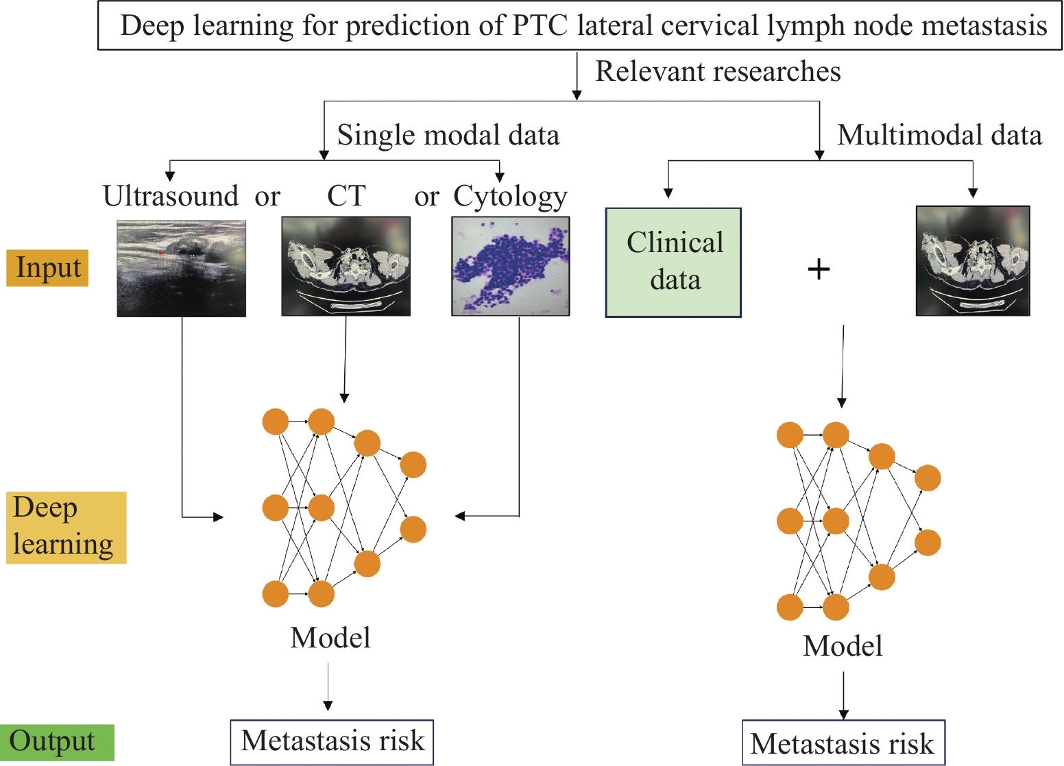
深度学习技术将传统医学图像转换为定量数据,从经验医学诊断走向数字医学诊断,还可以识别肉眼不能识别的肿瘤微观特征,在影像病理分割、诊断、转移风险评估、生存率和治疗反应的预测等各个领域的应用均引起了广泛重视,尤其是以图像诊断为主的甲状腺疾病领域。通过深度学习和数字图像处理技术,实现对影像或病理图像的自动化分析实现诊断是目前深度学习辅助医疗应用的主流方向。目前国内外基于深度学习模型判断甲状腺结节良恶性已较为成熟,国内部分医疗中心超声影像设备也安装了相关软件,除此之外,也有数个已注册的研究正在开展。PTC侧颈淋巴结转移对手术决策至关重要,然而,目前有关侧颈淋巴结转移的评估手段有限,这是PTC精准治疗过程中面对的挑战。深度学习图像分析为PTC侧颈淋巴结转移客观精确的评估提供了可能。本研究中我们回顾了已报道的有关基于影像或病理,包括超声、CT、细胞学及多模态数据的深度学习模型并进行了总结,见图2。基于超声的深度学习模型对PTC侧颈淋巴结转移评估的准确率可达92%以上[28],但也有不足70%的报道[22]。同时,深度学习在基于增强CT、细胞学图像预测淋巴结转移方面也取得了可观的成绩。多模态研究是目前的研究热点领域,有研究表明使用多模态数据无论是基于常规的分析技术还是基于深度学习的分析技术均具有优于单模态数据的预测效果[20,22]。然而,目前的多模态技术尚不成熟,医疗多维度复杂数据的处理技术仍有待提升。
![]() 图 2 目前有关深度学习预测PTC侧颈淋巴结转移的相关报道概括Figure 2 Summary of reports on deep learning in predicting lateral cervical lymph node metastasis in PTC
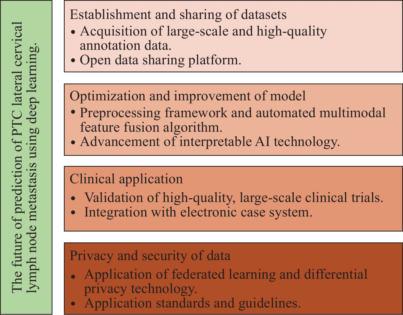
图 2 目前有关深度学习预测PTC侧颈淋巴结转移的相关报道概括Figure 2 Summary of reports on deep learning in predicting lateral cervical lymph node metastasis in PTC尽管深度学习在PTC淋巴结转移中的应用取得了诸多令人印象深刻的成果,但目前仍面临一些挑战。在数据层面,获取准确、全面且标注完善的大规模数据集一直是深度学习模型应用面临的巨大挑战,PTC原发灶和淋巴结影像或病理图像的标注需要专业人员进行,耗时耗力且成本高昂,这极大限制了大规模标注数据的获取。另外,数据不均衡以及医疗数据的隐私和安全问题也是影响模型鲁棒性及限制数据集规模和多样性的重要因素。模型的可解释性是另一个显著挑战,深度学习模型被视为“黑箱”结构,算法本身较关注精确度而忽略可解释性。可解释性在PTC侧颈淋巴结转移方面尤其重要,因为这与手术决策密切相关。虽然目前存在多种可视化技术以提高模型的可解释性,但可视化后的结果仍然难以完全解释。在模型的泛化能力方面,由于不同机构之间的影像设备、数据获取流程等存在差异,深度学习模型在一个机构的数据上训练后,往往难以在另一个机构上同样表现出色。最近来自于北卡罗来纳大学的研究发现,约43%FDA授权的人工智能医疗设备缺乏公开数据的验证[46],这也加重了临床工作者们对人工智能在医疗界应用的担忧。在计算资源方面,算力需求是目前面临的另一个显著挑战,基于CT或超声等二维图像的CNN模型通常也需要数十万乃至近千万的参数训练,即便是在计算机硬件快速发展的今天,深度学习模型的训练仍然需要花费大量的计算资源和时间,价格高昂的图形处理单元(GPU)或张量处理单元(TPU)设备也是限制深度学习广泛应用的重要因素。除此之外,由于医学伦理、法律与监管等问题,深度学习模型的临床试验及推广也是目前客观存在的困难。不过,目前深度学习仍在快速进展中,未来,随着大型数据集的建立与共享、模型算法的优化与改进、临床应用法律法规及数据隐私保护手段的完善,见图3,深度学习有可能成为医疗决策的重要参与者。
![]() 图 3 深度学习预测PTC侧颈淋巴结转移未来可能的方向Figure 3 Possible future of deep learning in predicting lateral cervical lymph node metastasis in PTC
图 3 深度学习预测PTC侧颈淋巴结转移未来可能的方向Figure 3 Possible future of deep learning in predicting lateral cervical lymph node metastasis in PTC深度学习在PTC侧颈淋巴结转移中的应用前景广阔,已在超声、CT、细胞学或多模态数据等方面展现出巨大潜力。然而,数据数量、质量、模型的解释性、多模态数据的处理、算法更新及伦理等问题仍需解决。随着更多高质量数据的积累、计算机技术的发展及法律法规的完善,深度学习有望构建更精确、更便捷的侧颈淋巴结转移预测模型,从而为手术决策和围术期咨询等方面提供更可靠的依据。
Competing interests: The authors declare that they have no competing interests.利益冲突声明:所有作者均声明不存在利益冲突。作者贡献:邵胜利:论文撰写、资料整理、项目负责人王吉恒:资料收集、论文撰写及审阅刘善廷:研究设计、立项负责及审阅 -
![]()
图 2 目前有关深度学习预测PTC侧颈淋巴结转移的相关报道概括
Figure 2 Summary of reports on deep learning in predicting lateral cervical lymph node metastasis in PTC
![]()
图 3 深度学习预测PTC侧颈淋巴结转移未来可能的方向
Figure 3 Possible future of deep learning in predicting lateral cervical lymph node metastasis in PTC
表 1 超声及CT在诊断PTC侧颈淋巴结转移中的表现
Table 1 Ultrasound and CT in diagnosis of lateral cervical lymph node metastasis in PTC
Literature Year Ultrasound CT Sample Sensitivity Specificity Sensitivity Specificity Ahn JE, et al [17] 2008 0.65 0.82 0.79 0.78 181 Kim E, et al[18] 2008 0.64 0.92 0.74 0.95 277 Choi JS, et al[19] 2009 0.94 0.25 0.82 1 352 Lee DW, et al[20] 2013 0.70 0.84 0.82 0.64 558 Lesnik D, et al[21] 2014 0.81 0.87 0.79 0.83 196 Na DK, et al[22] 2014 0.64 0.91 0.71 0.93 352 Lee Y, et al[23] 2018 0.74 0.90 0.82 0.90 764 Yang SY, et al[24] 2019 0.85 0.87 0.85 0.80 453  下载: 导出CSV
-
[1] 郑荣寿, 陈茹, 韩冰峰, 等. 2022年中国恶性肿瘤流行情况分析[J]. 中华肿瘤杂志, 2024, 46(3): 221-231. [Zheng RS, Chen R, Han BF, et al. Cancer incidence and mortality in China, 2022[J]. Zhonghua Zhong Liu Za Zhi, 2024, 46(3): 221-231.] doi: 10.3760/cma.j.cn112152-20240119-00035 Zheng RS, Chen R, Han BF, et al. Cancer incidence and mortality in China, 2022[J]. Zhonghua Zhong Liu Za Zhi, 2024, 46(3): 221-231. doi: 10.3760/cma.j.cn112152-20240119-00035
[2] Davies L, Welch HG. Current thyroid cancer trends in the United States[J]. JAMA Otolaryngol Head Neck Surg, 2014, 140(4): 317-322.
[3] 中国医师协会外科医师分会甲状腺外科医师委员会, 中国研究型医院学会甲状腺疾病专业委员会. 分化型甲状腺癌颈侧区淋巴结清扫专家共识(2017版)[J]. 中国实用外科杂志, 2017, 37(9): 985-991. [Chinese Thyroid Association, Chinese Collage of Surgeons, Chinese Medical Doctor Association, Thyroid Disease Professional Committee of Chinese Research Association. Expert Consensus on Cervical Lymph Node Dissection for Differentiated Thyroid Cancer (2017 Edition)[J]. Zhonghua Shi Yong Wai Ke Za Zhi, 2017, 37(9): 985-991.] Chinese Thyroid Association, Chinese Collage of Surgeons, Chinese Medical Doctor Association, Thyroid Disease Professional Committee of Chinese Research Association. Expert Consensus on Cervical Lymph Node Dissection for Differentiated Thyroid Cancer (2017 Edition)[J]. Zhonghua Shi Yong Wai Ke Za Zhi, 2017, 37(9): 985-991.
[4] 中国临床肿瘤学会指南工作委员会. 中国临床肿瘤学会(CSCO)分化型甲状腺癌诊疗指南2021[J]. 肿瘤预防与治疗, 2021, 34(12): 1164-1200. [Chinese Society of Clinical Oncology Guidance Working Committee. Chinese Society of Clinical Oncology (CSCO) Guidelines for The Diagnosis And Treatment of Differentiated Thyroid Cancer 2021[J]. Zhong Liu Yu Fang Yu Zhi Liao, 2021, 34 (12): 1164-1200.] Chinese Society of Clinical Oncology Guidance Working Committee. Chinese Society of Clinical Oncology (CSCO) Guidelines for The Diagnosis And Treatment of Differentiated Thyroid Cancer 2021[J]. Zhong Liu Yu Fang Yu Zhi Liao, 2021, 34 (12): 1164-1200.
[5] 中国抗癌协会甲状腺癌专业委员会, 中华医学会肿瘤学分会甲状腺肿瘤专业委员会, 中国研究型医院学会甲状腺疾病专业委员会, 等. 无充气腋窝入路腔镜甲状腺手术专家共识(2022版)[J]. 中华内分泌外科杂志, 2021, 15(6): 557-563. [Chinese Association of Thyroid Oncolog, Thyroid Tumor Committee of Oncology Branch of Chinese Medical Association, Thyroid Disease Professional Committee of Chinese Research Hospital Association, et al. Expert Consensus on Endoscopic Thyroidectomy by a Gasless Unilateral Axillary Approach (version 2022)[J]. Zhonghua Nei Fen Mi Wai Ke Za Zhi, 2021, 15(6): 557-563.] doi: 10.3760/cma.j.cn.115807-20211116-00349 Chinese Association of Thyroid Oncolog, Thyroid Tumor Committee of Oncology Branch of Chinese Medical Association, Thyroid Disease Professional Committee of Chinese Research Hospital Association, et al. Expert Consensus on Endoscopic Thyroidectomy by a Gasless Unilateral Axillary Approach (version 2022)[J]. Zhonghua Nei Fen Mi Wai Ke Za Zhi, 2021, 15(6): 557-563. doi: 10.3760/cma.j.cn.115807-20211116-00349
[6] Donangelo I, Walts AE, Bresee C, et al. Lymphocytic thyroiditis is associated with increased number of benign cervical nodes and fewer central neck compartment metastatic lymph nodes in patients with differentiated thyroid cancer[J]. Endocr Pract, 2016, 22(10): 1192-1198. doi: 10.4158/E151078.OR
[7] Uludag M, Cetinoglu I, Unlu MT, et al. The Role of Frozen Section Examination in Thyroid Surgery[J]. Sisli Etfal Hastan Tip Bul, 2023, 57(4): 441-450.
[8] Litjens G, Kooi T, Bejnordi BE, et al. A survey on deep learning in medical image analysis[J]. Med Image Anal, 2017, 42: 60-88. doi: 10.1016/j.media.2017.07.005
[9] Coudray N, Ocampo PS, Sakellaropoulos T, et al. Classification and mutation prediction from non-small cell lung cancer histopathology images using deep learning[J]. Nat Med, 2018, 24(10): 1559-1567. doi: 10.1038/s41591-018-0177-5
[10] Bilal M, Raza SEA, Azam A, et al. Development and validation of a weakly supervised deep learning framework to predict the status of molecular pathways and key mutations in colorectal cancer from routine histology images: a retrospective study[J]. Lancet Digit Health, 2021, 3(12): e763-e772. doi: 10.1016/S2589-7500(21)00180-1
[11] Kather JN, Pearson AT, Halama N, et al. Deep learning can predict microsatellite instability directly from histology in gastrointestinal cancer[J]. Nat Med, 2019, 25(7): 1054-1056.
[12] Yamashita R, Long J, Longacre T, et al. Deep learning model for the prediction of microsatellite instability in colorectal cancer: a diagnostic study[J]. Lancet Oncol, 2021, 22(1): 132-141. doi: 10.1016/S1470-2045(20)30535-0
[13] Sun Z, Zhang T, Ahmad MU, et al. Comprehensive assessment of immune context and immunotherapy response via noninvasive imaging in gastric cancer[J]. J ClinI Invest, 2024, 134(6): e175834.
[14] Xu Y, Hosny A, Zeleznik R, et al. Deep Learning Predicts Lung Cancer Treatment Response from Serial Medical Imaging[J]. Clin Cancer Res, 2019, 25(11): 3266-3275. doi: 10.1158/1078-0432.CCR-18-2495
[15] Yao J, Cao K, Hou Y, et al. Deep Learning for Fully Automated Prediction of Overall Survival in Patients Undergoing Resection for Pancreatic Cancer: A Retrospective Multicenter Study[J]. Ann Surg, 2023, 278(1): e68-e79. doi: 10.1097/SLA.0000000000005465
[16] Bera K, Braman N, Gupta A, et al. Predicting cancer outcomes with radiomics and artificial intelligence in radiology[J]. Nat Rev Clin Oncol, 2022, 19(2): 132-146. doi: 10.1038/s41571-021-00560-7
[17] Ahn JE, Lee JH, Yi JS, et al. Diagnostic accuracy of CT and ultrasonography for evaluating metastatic cervical lymph nodes in patients with thyroid cancer[J]. World J Surg, 2008, 32(7): 1552-1558. doi: 10.1007/s00268-008-9588-7
[18] Kim E, Park JS, Son KR, et al. Preoperative diagnosis of cervical metastatic lymph nodes in papillary thyroid carcinoma: comparison of ultrasound, computed tomography, and combined ultrasound with computed tomography[J]. Thyroid, 2008, 18(4): 411-418. doi: 10.1089/thy.2007.0269
[19] Choi JS, Kim J, Kwak JY, et al. Preoperative staging of papillary thyroid carcinoma: comparison of ultrasound imaging and CT[J]. AJR Am J Roentgenol, 2009, 193(3): 871-878.
[20] Lee DW, Ji YB, Sung ES, et al. Roles of ultrasonography and computed tomography in the surgical management of cervical lymph node metastases in papillary thyroid carcinoma[J]. Eur J Surg Oncol, 2013, 39(2): 191-196. doi: 10.1016/j.ejso.2012.07.119
[21] Lesnik D, Cunnane ME, Zurakowski D, et al. Papillary thyroid carcinoma nodal surgery directed by a preoperative radiographic map utilizing CT scan and ultrasound in all primary and reoperative patients[J]. Head Neck, 2014, 36(2): 191-202. doi: 10.1002/hed.23277
[22] Na DK, Choi YJ, Choi SH, et al. Evaluation of cervical lymph node metastasis in thyroid cancer patients using real-time CT-navigated ultrasonography: preliminary study[J]. Ultrasonography, 2015, 34(1): 39-44.
[23] Lee Y, Kim JH, Baek JH, et al. Value of CT added to ultrasonography for the diagnosis of lymph node metastasis in patients with thyroid cancer[J]. Head Neck, 2018, 40(10): 2137-2148.
[24] Yang SY, Shin JH, Hahn SY, et al. Comparison of ultrasonography and CT for preoperative nodal assessment of patients with papillary thyroid cancer: diagnostic performance according to primary tumor size[J]. Acta Radiol, 2020, 61(1): 21-27. doi: 10.1177/0284185119847677
[25] Lee JH, Baek JH, Kim JH, et al. Deep Learning-Based Computer-Aided Diagnosis System for Localization and Diagnosis of Metastatic Lymph Nodes on Ultrasound: A Pilot Study[J]. Thyroid, 2018, 28(10): 1332-1338.
[26] Zhou LQ, Zeng SE, Xu JW, et al. Deep learning predicts cervical lymph node metastasis in clinically node-negative papillary thyroid carcinoma[J]. Insights Imaging, 2023, 14(1): 222. doi: 10.1186/s13244-023-01550-2
[27] Zhao HN, Yin H, Liu JY, et al. Deep learning-assisted ultrasonic diagnosis of cervical lymph node metastasis of thyroid cancer: a retrospective study of 3059 patients[J]. Front Oncol, 2024, 14: 1204987. doi: 10.3389/fonc.2024.1204987
[28] Yuan Y, Pan B, Mo H, et al. Deep learning-based computer-aided diagnosis system for the automatic detection and classification of lateral cervical lymph nodes on original ultrasound images of papillary thyroid carcinoma: a prospective diagnostic study[J]. Endocrine, 2024, 85(3): 1289-1299. doi: 10.1007/s12020-024-03808-1
[29] Yuan Y, Hou S, Wu X, et al. Application of deep-learning to the automatic segmentation and classification of lateral lymph nodes on ultrasound images of papillary thyroid carcinoma[J]. Asian J Surg, 2024, 47(9): 3892-3898. doi: 10.1016/j.asjsur.2024.02.140
[30] Zhang MB, Meng ZL, Mao Y, et al. Cervical lymph node metastasis prediction from papillary thyroid carcinoma US videos: a prospective multicenter study[J]. BMC Med, 2024, 22(1): 153. doi: 10.1186/s12916-024-03367-2
[31] Yang J, Zhang F, Qiao Y. Diagnostic accuracy of ultrasound, CT and their combination in detecting cervical lymph node metastasis in patients with papillary thyroid cancer: a systematic review and meta-analysis[J]. BMJ Open, 2022, 12(7): e051568. doi: 10.1136/bmjopen-2021-051568
[32] Wang T, Yan D, Liu Z, et al. Diagnosis of cervical lymph node metastasis with thyroid carcinoma by deep learning application to CT images[J]. Front Oncol, 2023, 13: 1099104.
[33] Cho SJ, Suh CH, Baek JH, et al. Diagnostic performance of CT in detection of metastatic cervical lymph nodes in patients with thyroid cancer: a systematic review and meta-analysis[J]. Eur Radiol, 2019, 29(9): 4635-4647. doi: 10.1007/s00330-019-06036-8
[34] Suh CH, Baek JH, Choi YJ, et al. Performance of CT in the Preoperative Diagnosis of Cervical Lymph Node Metastasis in Patients with Papillary Thyroid Cancer: A Systematic Review and Meta-Analysis[J]. AJNR Am J Neuroradiol, 2017, 38(1): 154-161. doi: 10.3174/ajnr.A4967
[35] Zheng G, Zhang H, Lin F, et al. Performance of CT-based deep learning in diagnostic assessment of suspicious lateral lymph nodes in papillary thyroid cancer: a prospective diagnostic study[J]. Int J Surg, 2023, 109(11): 3337-3345.
[36] Lee JH, Ha EJ, Kim JH. Application of deep learning to the diagnosis of cervical lymph node metastasis from thyroid cancer with CT[J]. Eur Radiol, 2019, 29(10): 5452-5457.
[37] Lee JH, Ha EJ, Kim D, et al. Application of deep learning to the diagnosis of cervical lymph node metastasis from thyroid cancer with CT: external validation and clinical utility for resident training[J]. Eur Radiol, 2020, 30(6): 3066-3072. doi: 10.1007/s00330-019-06652-4
[38] Zhao W, Shen S, Ke T, et al. Clinical value of dual-energy CT for predicting occult metastasis in central neck lymph nodes of papillary thyroid carcinoma[J]. Eur Radiol, 2024, 34(1): 16-25.
[39] Chen M, Jiang Y, Zhou X, et al. Dual-Energy Computed Tomography in Detecting and Predicting Lymph Node Metastasis in Malignant Tumor Patients: A Comprehensive Review[J]. Diagnostics (Basel), 2024, 14(4): 377. doi: 10.3390/diagnostics14040377
[40] An C, Li D, Li S, et al. Deep learning radiomics of dual-energy computed tomography for predicting lymph node metastases of pancreatic ductal adenocarcinoma[J]. Eur J Nucl Med Mol Imaging, 2022, 49(4): 1187-1199. doi: 10.1007/s00259-021-05573-z
[41] Chung YJ, Lee JS, Park SY, et al. Histomorphological factors in the risk prediction of lymph node metastasis in papillary thyroid carcinoma[J]. Histopathology, 2013, 62(4): 578-588. doi: 10.1111/his.12025
[42] Ren W, Zhu Y, Wang Q, et al. Deep learning prediction model for central lymph node metastasis in papillary thyroid microcarcinoma based on cytology[J]. Cancer Sci, 2023, 114(10): 4114-4124. doi: 10.1111/cas.15930
[43] Lee Y, Alam MR, Park H, et al. Improved Diagnostic Accuracy of Thyroid Fine-Needle Aspiration Cytology with Artificial Intelligence Technology[J]. Thyroid, 2024, 34(6): 723-734. doi: 10.1089/thy.2023.0384
[44] Zhang Z, Zhang X, Yin Y, et al. Integrating BRAF(V600E) mutation, ultrasonic and clinicopathologic characteristics for predicting the risk of cervical central lymph node metastasis in papillary thyroid carcinoma[J]. BMC Cancer, 2022, 22(1): 461.
[45] Min Y, Huang Y, Wei M, et al. Preoperatively Predicting the Central Lymph Node Metastasis for Papillary Thyroid Cancer Patients With Hashimoto's Thyroiditis[J]. Front Endocrinol (Lausanne), 2021, 12: 713475. doi: 10.3389/fendo.2021.713475
[46] Chouffani El Fassi S, Abdullah A, Fang Y, et al. Not all AI health tools with regulatory authorization are clinically validated[J]. Nat Med, 2024, 30(10): 2718-2720. doi: 10.1038/s41591-024-03203-3

计量
- 文章访问数: 1258
- HTML全文浏览量: 5826
- PDF下载量: 840

