
Pathogenesis of Radioactive Esophagitis Based on TGF-β1/p38MAPK/FN Signaling Pathway
-
摘要:目的
从黏膜修复角度探讨放射性食管炎的发病机制,并明确是否与TGF-β1/p38MAPKs/FN信号通路相关。
方法HE染色法对放射性食管炎标本进行病理分析,Real-time PCR法检测标本FN、TGF-β1基因表达水平,Western blot法检测组织蛋白TGF-β1、p38、FN的表达。
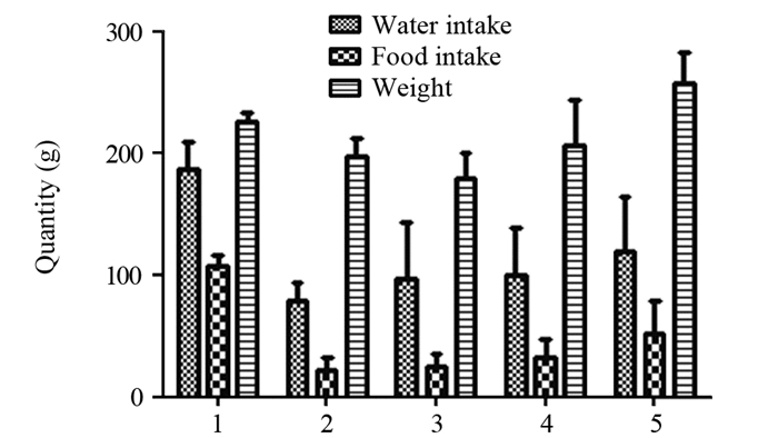
结果SD大鼠发生放射性食管炎后第一周体质量、进食量、进水量明显下降(P < 0.05),第四周恢复;病理方面,第一、二周食管黏膜层破坏,第四周出现再生;黏膜相关细胞因子TGF-β1、FN与病理变化一致,TGF-β1、p38MAPK蛋白表达先上升后下降,而FN蛋白表达先下降后上升。
结论TGF-β1/p38MAPK/FN信号通路可能参与了其黏膜修复过程。
-
关键词:
- TGF-β1/p38MAPK/FN信号通路 /
- 放射性食管炎 /
- 黏膜修复 /
- 细胞因子
Abstract:ObjectiveTo investigate the pathogenesis mechanism of radiation esophagitis from the perspective of mucosal regeneration and to determine whether it is associated with TGF-β1/p38MAPKs/FN signaling pathway.
MethodsThe pathological analysis of esophageal specimens was performed by HE staining method. The expression of FN and TGF-β1 genes were observed by real time-PCR method, and the expression of tissue proteins TGF-β1, p38 and FN were detected by Western blot.
ResultsThe weights, food intakes and water intakes at the first week after the occurrence of radiation esophagitis were significantly decreased (P < 0.05) and recovered at the fourth week. The esophageal mucosa was destructed at the first and second weeks, and the regeneration occurred in the fourth weeks; TGF-β1 and p38MAPK protein expression increased first and then decreased, while FN protein expression decreased first and then increased.
ConclusionThe TGF-β1/p38MAPK/FN signaling pathway may be involved in the process of mucosal repair.
-
Competing interests: The authors declare that they have no competing interests.作者贡献张福鹏:课题设计及文章撰写赵晓燕、郝淑兰、樊桂玲、王爱荣:实验设计与操作李晓丽:统计分析刘丽坤:文章指导
-
![]()
图 1 SD大鼠进水、进食量及体质量的动态变化
Figure 1 Dynamic changes of water intake, food intake and body weight of SD rats
![]()
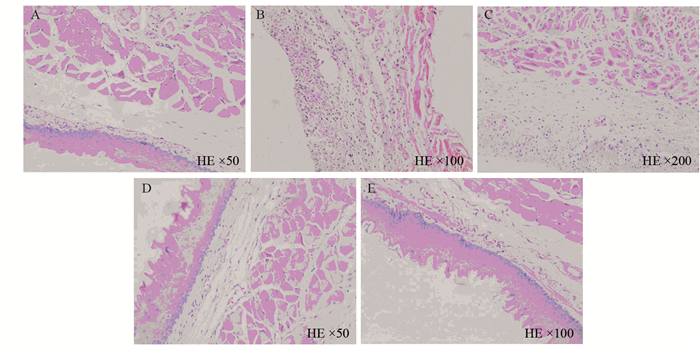
图 2 放射性食管炎SD模型食管病理的动态变化
Figure 2 Dynamic pathological changes of esophagus in SD model of radiation esophagitis
![]()
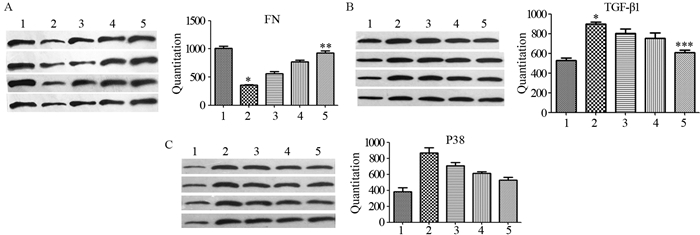
图 3 放射性食管炎SD大鼠模型食管组织FN(A)、TGF-β1(B)和P38(C)蛋白表达
Figure 3 Expression of FN(A), TGF-β1(B) and P38(C) protein in esophagus tissue of SD rat model of radiation esophagitis
表 2 放射性食管炎SD大鼠模型食管组织FN、TGF-β1 mRNA的表达
Table 2 Expression of FN and TGF-β1 mRNA in esophagus tissue of SD rat model of radiation esophagitis

 下载: 导出CSV
-
[1] Baker S, Fairchild A. Radiation-induced esophagitis in lung cancer[J]. Lung Cancer (Auckl), 2016, 7: 119-127.
[2] Wada K, Kishi N, Kanayama N, et al. Predictors of Acute Radiation Esophagitis in Non-small Cell Lung Cancer Patients Treated With Accelerated Hyperfractionated Chemoradiotherapy[J]. Anticancer Res, 2019, 39(1): 491-497. doi: 10.21873/anticanres.13139
[3] Pu X, Wang L, Chang JY, et al. Inflammation-related genetic variants predict toxicity following definitive radiotherapy for lung cancer[J]. Clin Pharmacol Ther, 2014, 96(5): 609-615. doi: 10.1038/clpt.2014.154
[4] Bowen JM, White I, Smith L, et al. Pre-therapy mRNA expression of TNF is associated with regimen-related gastrointestinal toxicity in patients with esophageal cancer: a pilot study[J]. Support Care Cancer, 2015, 23(11): 3165-3172. doi: 10.1007/s00520-015-2696-7
[5] Bosch DJ, Wang D, Nijsten MWN, et al. Longitudinal analysis of cytokine expression during neoadjuvant chemoradiotherapy and subsequent surgery in esophageal cancer patients[J]. Am J Surg, 2016, 212(1): 89-95. doi: 10.1016/j.amjsurg.2015.12.021
[6] 李婉迪, 赵振民.转化生长因子-β/Smads信号通路在病理性瘢痕形成中的作用机制研究进展[J].新乡医学院学报, 2017, 34(4): 335-339. http://www.cnki.com.cn/Article/CJFDTOTAL-XXYX201704026.htm Li WD, Zhao ZM. Research progress on the mechanism of transforming growth factor-β/Smads signaling pathway in pathological scar formation[J]. Xinxiang Yi Xue Yuan Xue Bao, 2017, 34(4): 335-339. http://www.cnki.com.cn/Article/CJFDTOTAL-XXYX201704026.htm
[7] D.L.斯托克姆, 庞希宁, 付小兵.再生生物学与再生医学[M].第1版.北京:科学出版社, 2013: 29-30. Stocum DL, Pang XN, Fu XB. Regenerative Biology and Medicine[M]. First edition. Beijing: Science Press, 2013: 29-30.
[8] Kwak EA, Lee NY. Synergetic roles of TGF-β signaling in tissue engineering[J]. Cytokine, 2019, 115: 60-63. doi: 10.1016/j.cyto.2018.12.010
[9] 于翔, 戴铭卉, 刘猛, 等.通腑泄浊法对慢性肾脏病大鼠TGF-β1/p38MAPK信号通路的调节作用[J].中成药, 2019, 41(5): 1000-1005. http://www.cnki.com.cn/Article/CJFDTotal-ZCYA201905009.htm Yu X, Dai MH, Liu M, et al. Regulatory effects of Discharging Fu-organ and Removing Turbidity Method on TGF-β1/p38MAPK signaling pathway in chronic kidney disease rats[J]. Zhong Cheng Yao, 2019, 41(5): 1000-1005. http://www.cnki.com.cn/Article/CJFDTotal-ZCYA201905009.htm
[10] 张兰, 刘晶, 刘天龙, 等.黄芪三萜皂苷通过抑制TGF-β1/Smad2、p38MAPK信号抑制CVB3诱导的心肌纤维化[J].中国分子心脏病学杂志, 2018, 18(5): 2631-2634. http://www.cnki.com.cn/Article/CJFDTotal-ZGFB201805011.htm Zhang L, Liu J, Liu TL, et al. Astragalus triterpenoid saponins attenuated CVB3-1nduced cardiac fibrosis via suppressed TGF-1/Smad2, p38MAPK signal pathway[J]. Zhongguo Fen Zi Xin Zang Bing Xue Za Zhi, 2018, 18(5): 2631-2634. http://www.cnki.com.cn/Article/CJFDTotal-ZGFB201805011.htm
[11] 路军章, 孙志高, 蒲香蓉, 等.构建6MV-X射线的大鼠放射性食管炎模型[J].解放军医学院学报, 2016, 37(2): 167-170. http://www.cnki.com.cn/Article/CJFDTotal-JYJX201602020.htm Lu JZ, Sun ZG, Pu XR, et al. Establishment of radioactive esophagitis rats models by 6MV X-ray energy[J]. Jie Fang Jun Yi Xue Yuan Xue Bao, 2016, 37(2): 167-170. http://www.cnki.com.cn/Article/CJFDTotal-JYJX201602020.htm
[12] Shimbori C, Bellaye PS, Xia J, et al. Fibroblast growth factor-1 attenuates TGF-β1-induced lung fibrosis[J]. J Pathol, 2016, 240(2): 197-210. doi: 10.1002/path.4768
[13] Bao C, Yang Z, Cai Q, et al. Incremental load training improves renal fibrosis by regulating the TGF-β1/TAK1/MKK3/p38MAPK signaling pathway and inducing the activation of autophagy in aged mice[J]. Int J Mol Med, 2019, 44(5): 1677-1686.
[14] Pejchal J, Novotný J, Mařák V, et al. Activation of p38 MAPK and expression of TGF-β1 in rat colon enterocytes after whole body γ-irradiation[J]. Int J Radiat Biol, 2012, 88(4): 348-358. doi: 10.3109/09553002.2012.654044
[15] 徐文燕, 管青聪, 徐芳.丹参提取物对系膜增生性肾小球肾炎大鼠TGF-β1/p38MAPK信号通路的影响研究[J].新中医, 2020, 52(2): 5-9. Xu WY, Guan QC, Xu F. Extract of Radix et Rhizoma Salviae Miltiorrhizae has Effect on TGF-β1/p38MAPK Signaling Pathways in Rats with Mesangial Proliferative Glomerulonephritis[J]. Xin Zhong Yi, 2020, 52(2): 5-9.
[16] Van JA, Scholey JW, Konvalinka A. Insights into Diabetic Kidney Disease Using Urinary Proteomics and Bioinformatics[J]. J Am Soc Nephrol, 2017, 28(4): 1050-1061. doi: 10.1681/ASN.2016091018
[17] Chin BY, Mohsenin A, Li SX, et al. Stimulation of pro-alpha(1)(Ⅰ) collagen by TGF-beta(1) in mesangial cells: role of the p38 MAPK pathway[J]. Am J Physiol Renal Physiol, 2001, 280(3): F495-F504. doi: 10.1152/ajprenal.2001.280.3.F495
[18] Obata T, Brown GE, Yaffe MB. MAP kinase pathways activated by stress: the p38 MAPK pathway[J]. Crit Care Med, 2000, 28(4 Suppl): N67-N77.
[19] 庄奇新, 孟令平.食管疾病影像学[M].上海:上海科学技术出版社, 2017: 4. Zhuang QX, Meng LP. Imaging of Esophageal Diseases[M]. Shanghai: Shanghai Science and Technology Publishing House, 2017: 4.

计量
- 文章访问数: 2083
- HTML全文浏览量: 490
- PDF下载量: 818

