
Effect of LncRNA-p21 Regulating Notch Signaling Pathway on Proliferation, Migration and Invasion of Non-small Cell Lung Cancer A549 Cells
-
摘要:目的
探讨LncRNA-p21调控Notch信号通路对非小细胞肺癌A549细胞增殖、迁移及侵袭的影响。
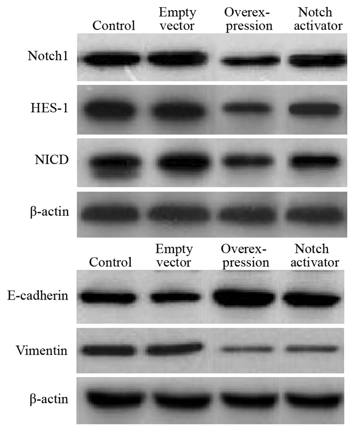
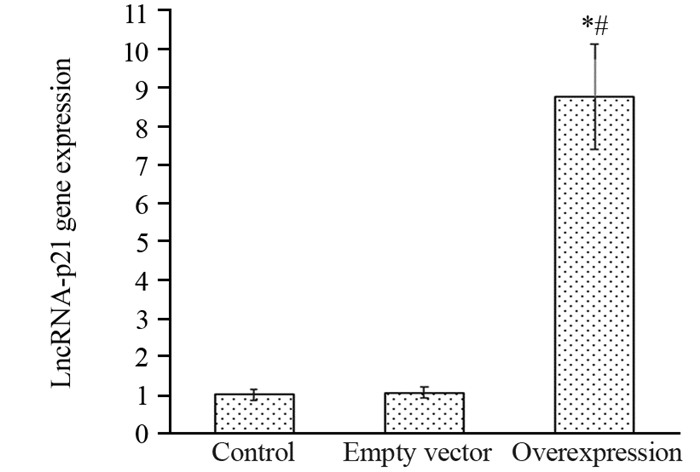
方法pcDNA-lincRNA-p21、空载质粒pcDNA转染A549细胞设为过表达组和空载组;稳定转染过表达组加入Notch信号通路特异性激活剂Jagged1蛋白,设为Notch激活剂组;不作处理细胞为对照组。MTT法、划痕实验和Transwell小室实验检测各组细胞增殖、迁移和侵袭情况。RT-qPCR及Western blot法检测各组Notch1、HES-1、NICD、E-cadherin、Vimentin的mRNA和蛋白表达。
结果过表达组培养24、48和72 h MTT实验A值均低于对照组、空载组和Notch激活剂组,Notch激活剂组低于对照组和空载组(P < 0.05);过表达组48 h细胞迁移率和穿膜细胞数及Notch1、HES-1、NICD、Vimentin mRNA和蛋白相对表达量均低于对照组、空载组和Notch激活剂组,Notch激活剂组低于对照组和空载组(P < 0.05);过表达组E-cadherin mRNA和蛋白相对表达量高于对照组和Notch激活剂组,Notch激活剂组高于空载组和对照组(P < 0.05)。
结论LncRNA-p21基因过表达可抑制非小细胞肺癌A549细胞增殖、迁移及侵袭,其调控机制可能与抑制Notch信号通路、阻断A549细胞上皮间质转化有关。
-
关键词:
- 非小细胞肺癌 /
- 长链非编码RNA-p21 /
- 增殖 /
- 迁移 /
- 侵袭
Abstract:ObjectiveTo investigate the effect of LncRNA-p21 on the proliferation, migration and invasion of non-small cell lung cancer A549 cells by regulating Notch signaling pathway.
MethodsThe pcDNA-lincRNA-p21 and empty plasmid pcDNA were transfected into A549 cells, and they were divided into overexpression group and empty vector group. Cells from the stably-transfected overexpression group were added with the Notch signaling pathway specific activator Jagged1 protein and set as the Notch activator group. In addition, the cells without treatment were taken as the control group. Cell proliferation, migration and invasion of each group were detected by MTT method, scratch test and Transwell cell test. The expressions of Notch 1, HES-1, NICD, E-cadherin, Vimentin mRNA and protein were detected by RT-qPCR and Western blot.
ResultsThe A value of MTT test at 24, 48 and 72 hours in the overexpression group was lower than those in the control group, empty vector group and Notch activator group, and the Notch activator group was higher than the control group and the empty vector group (P < 0.05). The cell migration rate, the number of transmembrane cells and the relative expressions of Notch1, HES-1, NICD, Vimentin mRNA and protein of overexpression group at 48 hours were lower than those of the control group, empty vector group and Notch activator group, and the Notch activator group was lower than the control group and the empty vector group (P < 0.05). The relative expressions of E-cadherin mRNA and protein of overexpression group were higher than those of control group, empty vector group and Notch activator group, and the Notch activator group was higher than the control group and the empty vector group (P < 0.05).
ConclusionOverexpression of LncRNA-p21 gene could inhibit the proliferation, migration and invasion of non-small cell lung cancer A549 cells. Its regulatory mechanism may be related to inhibiting Notch signaling pathway, thereby blocking the epithelial-mesenchymal transition of A549 cells.
-
Key words:
- Non-small cell lung cancer /
- Long non-coding RNA-p21 /
- Proliferation /
- Migration /
- Invasion
-
0 引言
目前,全球癌症的发病和死亡人数仍在持续增长,2020年新发肿瘤1 929.3万例、死亡995.8万例,癌症在许多国家的死因顺位上已经超越心血管疾病等高死亡率慢性疾病[1]。沈阳市城区居民全死因中,恶性肿瘤也仅次于心脏病[2],严重危害沈阳人民健康。为了解沈阳市城区居民恶性肿瘤发病及生存情况,并提出防治措施,现对2011—2018年沈阳市城区居民恶性肿瘤发病及生存趋势进行分析。
1 资料与方法
1.1 资料来源
2011—2018年沈阳市城区恶性肿瘤发病和生存资料来源于中国肿瘤登记中心肿瘤随访与登记报告系统。报告范围为具有沈阳市城市国家级监测点(和平区、沈河区、大东区、皇姑区和铁西区)户籍居民发病的全部恶性肿瘤(ICD10编码C00-C97)。人口资料来源于沈阳市公安局提供的每年份性别、年龄别的平均人口数。
1.2 质量评价
根据《中国肿瘤登记工作指导手册》[3]和国际癌症研究中心(IARC)/国际癌症登记协会(IACR)[4-5]对登记质量的有关要求,对数据的可靠性、完整性、有效性进行评估。沈阳市城区肿瘤发病数据从2008年起连续五年被国际五大洲癌症协会收录,连续十年被中国肿瘤登记年报收录。沈阳市城区2011—2018年上报新发肿瘤数据质量评价为病理诊断率(MV%)为66.63%、只有死亡医学证明书比例(DCO%)为2.43%、死亡发病比(M/I)为0.65,均符合质量要求。
1.3 统计学方法
提取2011—2018年沈阳城区恶性肿瘤发病数据,以国际疾病分类法ICD-10进行分类,并用IARCcrgTools软件进行数据审核。利用Excel2007计算粗发病率、标化率(中标率、世标率)、年龄别发病率、累积发病率(0~74岁)、截缩发病率(35~64岁)、前十位肿瘤发病顺位和生存率等指标;利用SPSS23.0统计软件对恶性肿瘤男女发病率及生存率进行χ2检验,检验水准α=0.05;采用寿命表法计算观察生存率,病例随访时间截止至2020-12-31;采用EdererⅡ方法(Ederer and Heise, 1959)计算期望生存率(expected survival rate, ESR)和相对生存率(relative survival rate, RSR);利用美国癌症中心研究所开发的Joinpoint 3.5.3软件计算发病率和生存率年度变化百分比(APC%),检验水准α=0.05。中标率采用2000年全国人口普查,世标率采用Segi's世界标准人口结构进行计算。
2 结果
2.1 总体发病分布
2011—2018年沈阳市上报新发恶性肿瘤109 873例,发病率为364.70/10万,中标率190.00/10万,世标率185.63/10万,0~74岁累积率为21.17%,35~64岁截缩率为311.66/10万。2011—2014年男、女、合计恶性肿瘤发病率及标化率均呈大幅上升趋势(P < 0.01);2015—2018年男、女、合计恶性肿瘤发病率缓慢上升但标化率缓慢下降(P≥0.05),见表 1。
表 1 2011—2018年沈阳市城区居民恶性肿瘤发病率(1/105)Table 1 Malignant tumor incidence of urban residents in Shenyang from 2011 to 2018 (1/105)
2.2 性别分布
2011—2018年沈阳市城区恶性肿瘤男性发病率为380.62/10万,女性发病率为349.42/10万,男、女发病率之比为1.09:1,8年间男性恶性肿瘤发病率高于女性,差异有统计学意义(χ2=201.63, P < 0.05),见表 1。
2.3 年龄分布
多数肿瘤在0~30岁组开始发病,30~40岁组缓慢上升,在40岁以后开始大幅度上升,在80~岁组达到发病高峰,85+岁组发病率略有下降,这可能与85+岁组人口急剧减少有关。男女以50~55岁组为界,发病率呈现X形状,见图 1。
![]() 图 1 2011—2018年沈阳市城区居民恶性肿瘤年龄别发病率Figure 1 Age-specific incidence of malignant tumor in urban areas of Shenyang city from 2011-2018
图 1 2011—2018年沈阳市城区居民恶性肿瘤年龄别发病率Figure 1 Age-specific incidence of malignant tumor in urban areas of Shenyang city from 2011-20182.4 发病顺位
2011—2018年沈阳市城区男性恶性肿瘤发病前10位的依次是肺癌、结直肠癌、肝癌、胃癌、膀胱癌、食管癌、胰腺癌、前列腺癌、肾癌、甲状腺癌;其中前5位肺癌(28.77%)、结直肠癌(16.40%)、肝癌(9.18%)、胃癌(9.15%)、膀胱癌(4.36%)占男性恶性肿瘤的67.86%。8年间肺癌、结直肠癌、膀胱癌、胰腺癌、前列腺癌、肾癌、甲状腺癌发病率均呈上升趋势(P < 0.05);而肝癌(P=0.00, P=0.05)、胃癌(P=0.02, P=0.07)、食管癌(P=0.08, P=0.24)发病率则先上升后下降,见表 2。
表 2 2011—2018年沈阳市城区男性恶性肿瘤发病顺位(1/105)Table 2 Incidence rank of malignant tumors in male residents in urban areas of Shenyang from 2011 to 2018 (1/105)
女性恶性肿瘤发病率前10位的依次是乳腺癌、肺癌、结直肠癌、宫颈癌、甲状腺癌、胃癌、肝癌、卵巢癌、胰腺癌、子宫体癌;其中前5位乳腺癌(22.63%)、肺癌(18.72%)、结直肠癌(12.84%)、宫颈癌(5.73%)、甲状腺癌(5.71%)占女性恶性肿瘤65.63%;8年间除宫颈癌、胃癌、肝癌、卵巢癌、子宫体癌外,乳腺癌、肺癌、结直肠癌、甲状腺癌、胰腺癌发病率均呈上升趋势(P < 0.05),见表 3。
表 3 2011—2018年沈阳市城区女性恶性肿瘤发病顺位(1/105)Table 3 Incidence rank of malignant tumors in female residents in urban areas of Shenyang from 2011 to 2018 (1/105)
2.5 恶性肿瘤5年生存率
2011—2015年沈阳市城区居民恶性肿瘤5年生存率为40.49%,相对生存率为47.84%。5年间合计观察生存率呈上升趋势,差异有统计学意义(P=0.04),其中男性为31.82%,女性为49.58%,男女观察生存率均呈上升趋势(P=0.04, P=0.03),且女性5年生存率高于男性(χ2=187.62, P < 0.05),见表 4。
表 4 2011—2015年沈阳市城区居民恶性肿瘤5年生存率(%)Table 4 Five-year survival rate of malignant tumors in urban residents in Shenyang from 2011 to 2015 (%)
2.6 发病前十位恶性肿瘤5年生存率顺位
2011—2015年沈阳市城区男性发病前十位的恶性肿瘤5年生存率顺位依次是甲状腺癌(86.25%)、肾癌(64.19%)、膀胱癌(59.43%)、结直肠癌(48.41%)、前列腺癌(47.55%)、胃癌(31.63%)、食管癌(20.56%)、肝癌(17.20%)、肺癌(16.79%)、胰腺癌(8.67%),见表 5。女性依次是甲状腺癌(91.81%)、乳腺癌(76.50%)、子宫体癌(73.17%)、子宫颈癌(65.18%)、结直肠癌(49.04%)、卵巢癌(43.34%)、胃癌(32.47%)、肺癌(21.20%)、肝癌(14.41%)、胰腺癌(10.01%),见表 6。
表 5 2011—2015年沈阳市城区男性发病前十位恶性肿瘤5年生存率(%)Table 5 Five-year survival rate of top ten malignant tumors among males in urban areas of Shenyang from 2011 to 2015 (%) 表 6 2011—2015年沈阳市城区女性发病前十位恶性肿瘤5年生存率(%)Table 6 Five-year survival rate of top ten malignant tumors among females in urban areas of Shenyang from 2011 to 2015 (%)
男女5年生存率最高均为甲状腺癌,最低均为胰腺癌。相同癌种中,肺癌、甲状腺癌5年生存率女性高于男性(χ2=48.29, χ2=9.85, P < 0.01),差异有统计学意义;肝癌男性高于女性(χ2=5.32, P < 0.05),差异有统计学意义;结直肠癌(χ2=0.37, P≥0.05)、胃癌(χ2=0.33, P≥0.05)、胰腺癌(χ2=0.99, P≥0.05)男女5年生存率差异无统计学意义。
2.7 发病前十位恶性肿瘤5年生存率变化趋势
2011—2015年沈阳市男性发病前十位的恶性肿瘤除结直肠癌、前列腺癌、肝癌外,其余七位中,甲状腺癌(APC%=12.97, P=0.03)、肾癌(APC%=7.86, P=0.01)、膀胱癌(APC%=10.04, P=0.00)、胃癌(APC%=6.57, P=0.05)、食管癌(APC%=6.05, P=0.03)、肺癌(APC%=11.81, P=0.04)、胰腺癌(APC%=25.57, P=0.02)5年生存率均呈上升趋势,见表 5。
女性发病前十位的恶性肿瘤除子宫体癌、结直肠癌、胃癌、肺癌外,其余六位的甲状腺癌(APC%=7.93, P=0.01)、乳腺癌(APC%=3.87, P=0.05)、宫颈癌(APC%=4.96, P=0.00)、卵巢癌(APC%=10.75, P=0.03)、肝癌(APC%=20.09, P=0.01)、胰腺癌(APC%=49.75, P=0.01)5年生存率均呈上升趋势,见表 6。
3 讨论
从发病情况看,沈阳市2011—2018年城区居民恶性肿瘤发病率持续上升(P=0.00, P=0.67),发病中标率(190.00/10万)低于2015年中国城市恶性肿瘤发病中标率(193.93/10万)[6]和中国东部恶性肿瘤发病中标率(194.36/10万)[7],高于中部恶性肿瘤发病中标率(183.36/10万)[8],低于2006—2015年辽宁省五城市恶性肿瘤标化发病率(199.15/10万)[9],高于2016年安徽(179.70/10万)[10]和江苏(182.61/10万)[11]、2017年黑龙江(174.27/10万)[12],目前发病率处于全国中等水平。
2011—2018年沈阳市恶性肿瘤发病率男性高于女性(χ2=201.63, P < 0.05),与中国分布相一致[13],且随着年龄的增长呈明显上升趋势,而沈阳市老龄人口亦呈逐年上升趋势,这既反映了人口老龄化进程的加快,也反映了癌症相关危险因素暴露时间的增加[14]是沈阳市癌症高发的重要原因。因此要针对病因和危险因素,致力于通过精准、适度和有效的干预,降低癌症发生风险。
2011—2018年男女恶性肿瘤发病顺位前三位与国家《2018肿瘤登记年报》发布中国城市数据[8]一致。8年间,男性发病前十位除肝癌、胃癌、食管癌发病率先上升后下降,其他癌种均呈上升趋势;女性除了妇科肿瘤和肝癌、胃癌外,其他发病前十的恶性肿瘤发病率均呈上升趋势。研究表明与感染或贫困相关的癌症正逐渐被经济发达国家的常见癌症所取代[15]。而沈阳市由于经济的发展和生活水平的提高,不良生活方式的加剧,不仅原有主要高发癌症尚未有明显下降趋势,西方国家高发的大肠癌、前列腺癌和女性乳腺癌等癌症发病又迅速增加。
从生存情况看,2011—2015年沈阳市城区恶性肿瘤5年观察生存率为40.49%、相对生存率为47.84%,接近于2018年全国公布的全部癌症的5年生存率(40.5%)[16],2019年辽宁省公布的城市癌症5年标化生存率(41.5%)[17],2012—2016年上海市青浦区5年相对生存率为47.41%[18],高于姑苏区2008—2013年癌症患者的5年相对生存率(42.2%)[19]。女性的生存率总体高于男性(χ2=187.62, P < 0.05)。
随着医疗技术水平的提升,沈阳市癌症5年生存率也大幅上升,但仍有部分癌种5年生存率无上升趋势,这与癌症种类构成不同和筛查手段落后造成生存率差异有重要关系。2011—2015年沈阳市城区恶性肿瘤5年生存率最高的癌种为甲状腺癌,其次是女性的乳腺癌(76.50%)、子宫体癌(73.17%)、宫颈癌(65.18%);男女生存率排在后三位是肝癌(17.20%、14.41%)、肺癌(16.79%、21.20%)、胰腺癌(8.67%、10.01%);生存率最低的均为胰腺癌。而这些生存率较高的癌症可以通过早期筛查项目,寻找出高危人群或早期患者,进行早发现、早诊断和早治疗的“二级预防”,是有效提升癌症生存的关键手段;对于生存率较低的癌症我们要依托于生物医学各学科不断发展的各种新技术、新手段,不断探索癌症相关标志物,优化筛查策略,早期发现、规范治疗,以提升患者生存率。
Competing interests: The authors declare that they have no competing interests.作者贡献:张冠磊:文献收集整理、实验设计及操作、统计分析、论文撰写与修改马苗苗:收集和资料汇总分析兰文静:文章审校、修改和指导王琳:论文指导 -
![]()
图 1 各组细胞中LncRNA-p21基因表达比较
Figure 1 Comparison of LncRNA-p21 gene expression in each group
![]()
图 2 各组划痕实验48 h细胞迁移率结果
Figure 2 Cell migration rates of each group at 48 hours detected by scratch test
![]()
图 3 Transwell实验48 h穿膜细胞数结果
Figure 3 Number of transmembrane cells at 48 hours detected by Transwell test
表 1 Notch1、HES-1、NICD、E-cadherin、Vimentin基因引物序列
Table 1 Primer sequences of Notch1, HES-1, NICD, E-cadherin and Vimentin genes

 下载: 导出CSV
表 2 各组细胞不同时间MTT试验A值比较
Table 2 Comparison of A value of MTT test at different time among each group
 下载: 导出CSV
表 3 Notch1、HES-1、NICD、E-cadherin和Vimentin mRNA相对表达量比较
Table 3 Comparison of relative expressions of Notch1, HES-1, NICD, E-cadherin and Vimentin mRNA
 下载: 导出CSV
表 4 Notch1、HES-1、NICD、E-cadherin和Vimentin蛋白相对表达量比较
Table 4 Comparison of relative expressions of Notch1, HES-1, NICD, E-cadherin and Vimentin protein
 下载: 导出CSV
-
[1] 季新强. 北京大学肿瘤医院2000-2013年3733例肺癌手术患者临床病理特征动态变化分析[J]. 中华肿瘤防治杂志, 2017, 24(24): 1687-1692. https://www.cnki.com.cn/Article/CJFDTOTAL-QLZL201724001.htm Ji XQ. Dynamic analysis of clinical epidemiology and the distribution of pathology types of patients with primary lung cancer in Peking University Cancer Hospital from 2000 to 2013[J]. Zhonghua Zhong Liu Fang Zhi Za Zhi, 2017, 24(24): 1687-1692. https://www.cnki.com.cn/Article/CJFDTOTAL-QLZL201724001.htm
[2] Sakin A, Sahin S, Atci MM, et al. The effect of different treatment modalities on survival in elderly patients with locally advanced non-small cell lung cancer[J]. Pulmonology, 2019, pii: S2531-0437(19)30215-6.
[3] Niazi Z, Garazhian E, Esfandi F, et al. Expression Analysis of the Long Non-Coding RNA LINC01433 in Lung Cancer[J]. Klin Onkol, 2019, 32(6): 453-455.
[4] Chen X, Shi Y, Zhou K, et al. A bibliometric analysis of long non-coding RNA and chemotherapeutic resistance research[J]. Oncotarget, 2019, 10(35): 3267-3275. doi: 10.18632/oncotarget.26938
[5] 裴洪利, 白尚星. 过表达lncRNA-p21通过介导Wnt/β-catenin信号通路抑制胃癌MGC-803细胞的生长与转移[J]. 临床与病理杂志, 2019, 39(8): 1615-1621. https://www.cnki.com.cn/Article/CJFDTOTAL-WYSB201908003.htm Pei HL, Bai SX. Over-expression of lncRNA-p21 inhibits the growth and metastasis of gastric cancer MGC-803 cells by mediating Wnt/beta-catenin signaling pathway[J]. Lin Chuang Yu Bing Li Za Zhi, 2019, 39(8): 1615-1621. https://www.cnki.com.cn/Article/CJFDTOTAL-WYSB201908003.htm
[6] Guo L, Gu J, Hou S, et al. Long non-coding RNA DANCR promotes the progression of non-small-cell lung cancer by inhibiting p21 expression[J]. Onco Targets Ther, 2018, 12: 135-146. doi: 10.2147/OTT.S186607
[7] Dahariya S, Paddibhatla I, Kumar S, et al. Long non-coding RNA: Classification, biogenesis and functions in blood cells[J]. Mol Immunol, 2019, 112: 82-92. doi: 10.1016/j.molimm.2019.04.011
[8] 张勇, 张启发, 田长海, 等. 长链非编码RNA-p21通过调节Warburg效应促进前列腺癌细胞侵袭[J]. 中华实验外科杂志, 2017, 34(6): 1014-1016. Zhang Y, Zhang QF, Tian CH, et al. Long non-coding RNA-p21 induced by hypoxia promotes the invasion of prostate cancer by enhancing Warburg effect[J]. Zhonghua Shi Yan Wai Ke Za Zhi, 2017, 34(6): 1014-1016.
[9] 朱克祥, 张正聪, 袁得峰, 等. lincRNA-p21通过STAT3信号抑制结直肠癌HCT116细胞增殖[J]. 中国病理生理杂志, 2019, 35(5): 797-803. https://www.cnki.com.cn/Article/CJFDTOTAL-ZBLS201905005.htm Zhu KX, Zhang ZC, Yuan DF, et al. Growth inhibition of colorectal cancer HCT116 cells by lincRNA-p21 through STAT3 signaling pathway[J]. Zhongguo Bing Li Sheng Li Za Zhi, 2019, 35(5): 797-803. https://www.cnki.com.cn/Article/CJFDTOTAL-ZBLS201905005.htm
[10] Zhou P, Wang C, Hu Z, et al. Genistein induces apoptosis of colon cancer cells by reversal of epithelial-to-mesenchymal via a Notch1/NF-κB/slug/E-cadherin pathway[J]. BMC Cancer, 2017, 17(1): 813. doi: 10.1186/s12885-017-3829-9
[11] Fukusumi T, Guo TW, Sakai A, et al. The NOTCH4-HEY1 Pathway Induces Epithelial-Mesenchymal Transition in Head and Neck Squamous Cell Carcinoma[J]. Clin Cancer Res, 2018, 24(3): 619-633. doi: 10.1158/1078-0432.CCR-17-1366
[12] Li N, Shi R. Expression alteration of long non-coding RNAs and their target genes in the intestinal mucosa of patients with Crohn's disease[J]. Clin Chim Acta, 2019, 494: 14-21. doi: 10.1016/j.cca.2019.02.031
[13] Han W, Liu J. LncRNA-p21 inhibited the proliferation of osteosarcoma cells via the miR-130b/PTEN/AKT signaling pathway[J]. Biomed Pharmacother, 2018, 97: 911-918. doi: 10.1016/j.biopha.2017.11.014
[14] Işın M, Uysaler E, Özgür E, et al. Exosomal lncRNA-p21 levels may help to distinguish prostate cancer from benign disease[J]. Front Genet, 2015, 6: 168.
[15] 张欣, 林雨, 海龙, 等. NOTCH通路依赖PI3K/AKT通路调控胶质瘤干细胞增殖和自我更新能力[J]. 肿瘤防治研究, 2018, 45(9): 640-646. doi: 10.3971/j.issn.1000-8578.2018.18.0029 Zhang X, Lin Y, Hai L, et al. NOTCH Signaling Pathway Regulates Glioma Stem-like Cell Proliferation and Self-renewal Abilities via PI3K/AKT Activity[J]. Zhong Liu Fang Zhi Yan Jiu, 2018, 45(9): 640-646. doi: 10.3971/j.issn.1000-8578.2018.18.0029
[16] Vázquez-Ulloa E, Ramos-Cruz AC, Prada D, et al. Loss of nuclear NOTCH1, but not its negative regulator NUMB, is an independent predictor of cervical malignancy[J]. Oncotarget, 2018, 9(27): 18916-18928. doi: 10.18632/oncotarget.24828
[17] Jiang B, Chen J, Yuan W, et al. Platelet-derived growth factor-D promotes colorectal cancer cell migration, invasion and proliferation by regulating Notch1 and matrix metalloproteinase-9[J]. Oncol Lett, 2018, 15(2): 1573-1579.
[18] 李美玲, 陈美琼, 张鹏, 等. 靶向沉默Notch1基因对骨髓瘤细胞增殖的影响[J]. 中国实验血液学杂志, 2017, 25(6): 1707-1712. https://www.cnki.com.cn/Article/CJFDTOTAL-XYSY201706027.htm Li ML, Chen MQ, Zhang P, et al. Targeting Notch1 Gene Inhibits the Proliferation of Multiple Myeloma Cells[J]. Zhongguo Shi Yan Xue Ye Xue Za Zhi, 2017, 25(6): 1707-1712. https://www.cnki.com.cn/Article/CJFDTOTAL-XYSY201706027.htm
[19] Pizza FX, Martin RA, Springer EM, et al. Intercellular adhesion molecule-1 augments myoblast adhesion and fusion through homophilic trans-interactions[J]. Sci Rep, 2017, 7(1): 5094.
[20] Zhang HS, Zhang ZG, Du GY, et al. Nrf2 promotes breast cancer cell migration via up-regulation of G6PD/HIF-1α/Notch1 axis[J]. J Cell Mol Med, 2019, 23(5): 3451-3463.
[21] Gao YP, Li Y, Li HJ, et al. LncRNA NBR2 inhibits EMT progression by regulating Notch1 pathway in NSCLC[J]. Eur Rev Med Pharmacol Sci, 2019, 23(18): 7950-7958.
-
期刊类型引用(3)
1. 贺明,汤成,梁艳,黄义娟,陶然. 2014-2016年重庆市九龙坡区新发恶性肿瘤生存分析. 社区医学杂志. 2023(03): 115-118+123 .  百度学术
百度学术
2. 李红,徐幽琼,郑婉辉,陆璐. 福州市2018年恶性肿瘤发病与死亡分析. 现代肿瘤医学. 2023(18): 3481-3485 . 百度学术
3. 亓琳,张欢欢,沈自芳,杨蕊. 彩色多普勒超声特征对乳腺无症状炎性改变和浸润性导管癌的鉴别诊断价值. 中国医药导报. 2023(28): 164-167 . 百度学术
其他类型引用(0)


计量
- 文章访问数: 2470
- HTML全文浏览量: 694
- PDF下载量: 406
- 被引次数: 3

