肿瘤防治研究  2014, Vol. 41 Issue (12): 1307-1312
本刊由国家卫生和计划生育委员会主管,湖北省卫生厅、中国抗癌协会、湖北省肿瘤医院主办。
0

文章信息

王玉祥,田丹丹,邱嵘,田秀明,王丽丽. 2014.
WANG Yuxiang, TIAN Dandan, QIU Rong, TIAN Xiuming, WANG Lili. 2014.
Ⅲ期非小细胞肺癌三维适形放疗所致放射性肺炎的影响因素
Influence Factors for Radiation Pneumonitis in Stage Ⅲ Non-small Cell Lung Cancer Patients Treated with Three-dimensional Conformal Radiotherapy
肿瘤防治研究, 2014, 41(12): 1307-1312
Cancer Research on Prevention and Treatment, 2014, 41 (12): 1307-1312
http://www.zlfzyj.com/CN/10.3971/j.issn.1000-8578.2014.12.012

文章历史

收稿日期:2013-11-13
修回日期:2014-07-01
Ⅲ期非小细胞肺癌三维适形放疗所致放射性肺炎的影响因素
王玉祥, 田丹丹, 邱嵘, 田秀明, 王丽丽    
050011 石家庄,河北医科大学第四医院放疗科(*:现单位:061001 沧州, 河北省沧州市中西医结合医院放疗科)
摘要目的 回顾性分析Ⅲ期非小细胞肺癌(NSCLC)三维适形放疗(3D-CRT)引起放射性肺炎(RP)发生的相关物理参数及临床影响因素。方法 2001年1月至2008年12月接受根治性3D-CRT的203例NSCLC临床资料,其中男163例、女40例;中位年龄63岁(30~83岁);21例有慢性阻塞性肺疾病(COPD)病史;ⅢA期79例、ⅢB期124例。70例单纯放疗、133例放化联合治疗,中位等效照射剂量6 200 cGy(5 000~7 800cGy);RP评价采用RTOG标准,剂量体积直方图(DVH)评价物理参数与≥2级和≥3级RP的关系。统计分析采用SPSS13.0统计软件。结果 203例放疗后发生≥2级RP者32%(65/203),≥3级RP者20.7%(42/203)。Spearman相关和Logistic单因素分析显示,物理参数中肺平均剂量、双肺V5~V40,临床因素中COPD病史、分割方式(常规分割/大分割)、放疗剂量、GTV和GTV/肺体积均与≥2级和≥3级RP相关(P<0.05);而性别、年龄、吸烟、病变部位(中央型/周围型、肺上叶/肺中下叶)、化疗与否等均与≥2级和≥3级RP无明显关系(P>0.05)。Logistic多因素回归分析显示,肺V25是≥2级和≥3级RP的独立危险因素,ROC曲线显示肺V25分界值为29%;COPD是≥2级RP的独立危险因素。结论 DVH参数可用于预测和评价放射性肺炎,肺V25可能是最有效预测≥2级RP和≥3级RP的指标,合并COPD增加≥2级RP的发生率。
关键词Ⅲ期非小细胞肺癌     三维适形放疗     放射性肺炎    
Influence Factors for Radiation Pneumonitis in Stage Ⅲ Non-small Cell Lung Cancer Patients Treated with Three-dimensional Conformal Radiotherapy
WANG Yuxiang, TIAN Dandan, QIU Rong, TIAN Xiuming, WANG Lili    
Department of Radiation Oncology, The Fourth Hospital of Hebei Medical University, Shijiazhuang 050011, China( *:Present: Department of Radiation Oncology, Cangzhou Hospital of Integrated Traditional Chinese and Western Medicine of Hebei Province,Cangzhou 061001, China)
AbstractObjective To retrospectively analyze physical parameters and clinical factors for radiation pneumonitis(RP) in stage Ⅲnon-small cell lung cancer(NSCLC) patients treated with three-dimensional conformal radiotherapy(3D-CRT). Methods From January 2001 to December 2008, 203 NSCLC patients were treated with radical 3D-CRT, 163 males and 40 females; Median ages was 63-year-old(30~83); 21 patients were suffered from chronic obstructive pulmonary disease(COPD) medical history; 79 cases were in ⅢA stage and 124 cases were in ⅢB stage; 70 cases were treated with single radiotherapy and 133 cases with chemoradiotherapy, and median equivalent dose was 6 200 cGy (5 000-7 800 cGy). RP was evaluated with RTOG standard, and physical parameters of DVH was used to estimated RP grade≥2 and RP grade≥ 3. SPSS 13.0 software was used for statistic analysis. Results After 3DCRT, the rates of RP grade≥2 and grade≥3 were 32%(65/203) and 20.7%(42/203). Spearman correlation and single factor Logistic analysis showed that the mean lung dose, V5-V40 of total lung, COPD history, fractional radiation style, radiotherapy dose, GTV and GTV/volume of total lung were correlated with RP grade≥2 and grade≥3 (P<0.05); Logistic multivariate analysis revealed that V25 was the independent risk factor for RP grade ≥2 and grade ≥3. The optimal cutoff value for lung V25 was 29% in the receiver-operating characteristic (ROC) curve. COPD was the independent risk factor for RP grade ≥2. Conclusion DVH parameters were associated with the occurrence of RP. V25 might be the most effectively independent risk factor for RP grade≥2 and grade ≥3. COPD was the independent risk factor for RP grade≥2 in patients with non-small cell lung cancer.
Key words: Stage Ⅲnon-small cell lung cancer     Three-dimensional conformal radiotherapy(3D-CRT)     Radiation pneumonitis(RP)    
0 引言

放疗是Ⅲ非小细胞肺癌(non-small cell lung cancer,NSCLC)主要治疗方法之一,放疗引起的放射性肺炎(尤其有症状放射性肺炎)是影响治疗疗效和生存质量的重要剂量限制性因素之一[1,2]。目前三维适形放疗(three-dimensional conformal radiotherapy,3D-CRT)已成为NSCLC的主要治疗技术,其放疗计划系统中剂量体积直方图(DVH)提供的物理参数,可以评价肺组织的受照剂量,通过优选比较,探寻出肺组织的可耐受剂量和有效预测放射性肺炎的参数和方法。为此,我们回顾性分析203例行3D- CRT的Ⅲ期NSCLC临床资料,评价有症状放射性肺炎(≥2级和≥3级)的相关物理参数和临床因素,便于指导临床治疗。1 资料与方法1.1 一般临床资料

收集2001年1月至2008年12月在河北医科大学第四医院放疗科行根治性三维适形放疗的203例Ⅲ期NSCLC临床资料,所有病例均有完整的放疗计划。其中男163例、女40例,中位年龄63岁(30~83岁);吸烟者133例、未吸烟者70例。21例有慢性阻塞性肺疾病(chronic obstructive pulmonary disease,COPD)、182例无COPD; 101例无阻塞性肺炎、102例有肺叶或肺段的阻塞性肺炎;原发肿瘤位于中央型173例、周围型30例;病变位于肺上叶(包括右肺中叶)111例、92例位于肺下叶。按照2003年UICC分期标准,ⅢA期79例、ⅢB期124例。1.2 治疗方法

3D-CRT采用热塑膜固定体位,CT模拟扫描定位,3D-CRT计划系统进行图象数字化传输、三维重建,勾画GTV包括原发病灶及转移淋巴结,将GTV均匀外扩6~8 mm为临床靶区(CTV),CTV外扩5~10 mm为计划靶区(PTV)。前程常规放疗者于模拟机下定位,照射野包括原发灶、转移淋巴结及相应淋巴引流区的亚临床灶,最后将前程常规照射野拟合到后程3D-CRT计划中进行评价。

203例中,44例采用前程常规放疗+后程三维适形放疗,126例采用全程三维适形放疗,33例采用调强放疗。21例采用大分割,2.5~4 Gy/f,182例采用常规分割,1.8~2.0 Gy/f;中位等效照射剂量6 200 cGy(范围5 000~7 800 cGy)。70例采用单纯放疗,133例放化联合治疗(序贯放化疗46例、同期放化疗87例),化疗方案有CAP(环磷酰胺+阿霉素+顺铂)、CFP(环磷酰胺+氟尿嘧啶+顺铂)、EP(依托泊苷+顺铂)、NP(长春瑞滨+顺铂)等;化疗周期数1~10个,中位数2周期。1.3 物理参数评价

根据DVH计算GTV、PTV、GTV和PTV最大剂量、双肺体积(指双肺体积-PTV后的体积)、双肺平均剂量(MLD),双肺V5、V10、V15、V20、V25、V30、V35、V40 是指双肺(双肺体积-PTV)接受5、10、15、20、25、30、35及40 Gy以上剂量照射的体积百分比。1.4 放射性肺炎评价标准

肺急性不良反应按照WHO早期反应评价标准和美国肿瘤放射治疗协作组(RTOG)急性放射损伤分级标准进行评价,放疗开始后3月内进行评价。1.5 统计学方法

采用SPSS13.0软件统计分析,计数资料采用χ2检验,变量间的相关性采用Spearman相关性检验,Logistic逐步回归分析筛选放射性肺炎的影响因素;受试者工作特征(receiver operating characteristic,ROC)曲线算出不同临界点时曲线下的面积(area under the curve,AUC)、敏感度、特异性,计算最佳临界值。P<0.05为差异具有统计学意义。2 结果2.1 放射性肺炎发生的相关因素

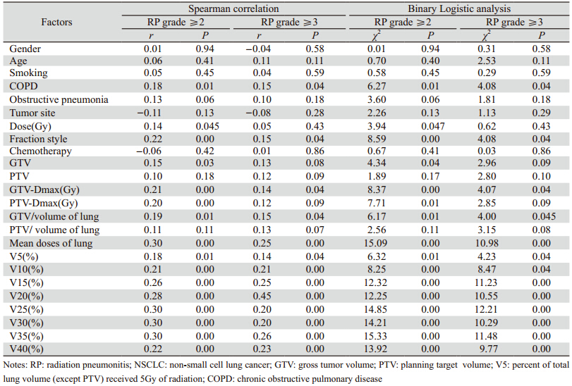
203例Ⅲ期NSCLC行3D-CRT后,发生0~1、2、3、4级放射性肺炎(RP)者分别为138、23、37、5例,≥2级RP者32.0%(65/203)、≥3级RP者20.7%(42/203)。Spearman相关和Binary Logistic单因素分析显示:COPD病史、分割方式(常规分割/大分割)、放疗剂量、物理参数中肺平均剂量(MLD)、肺V5~V40、GTV最大剂量、GTV/肺体积均与≥2级和≥3级RP相关(P<0.05);GTV、PTV最大剂量与≥2级RP相关(P<0.05),与≥3级RP也有相关性,但P>0.05;而性别、年龄、吸烟、病变部位(中央型/周围型、肺上叶/肺中下叶)、是否合并阻塞性肺炎、化疗与否、肺体积、PTV/肺体积等均与≥2级和≥3级RP无明显相关性(P>0.05),见表 1

表 1 Ⅲ期非小细胞肺癌患者放射性肺炎的单因素分析 Table 1 Univariate analysis of influence factors for RP in stage ⅢNSCLC patients
2.2 放射性肺炎发生的Logistic多因素分析

Spearman相关和Binary Logistic多因素回归分析,结果显示双肺V25是≥2级和≥3级RP的最佳预测指标,合并COPD是预测≥2级RP的独立预测指标,见表 2

表 2 Ⅲ期非小细胞肺癌患者放射性肺炎发生的Logistic多因素分析 Table 2 Multivariate logistic regression analysis of influence factors for RP in stage Ⅲ NSCLC patients
2.3 ROC曲线分析各指标预测放射性肺炎的效能

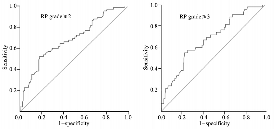
ROC曲线计算出预测≥2级和≥3级RP的双肺平均剂量、双肺V5、V10、V15、V20、V25、V30、V35、V40的分界值分别为1 840 cGy、64%、49%、36%、31%、29%、24%、20%和20%(P<0.05);其中V25预测≥2级和≥3级RP的敏感度、特异性、曲线下面积分别为67.6%和81.9%、54.8%和52.3%、0.685(0.605~0.764)和0.675(0.584~0.766),见图 1

ROC: receiver operating characteristic 图 1 Ⅲ期非小细胞肺癌患者肺V25诊断预测放射性肺炎的ROC曲线(误判率=1-特异性) Figure 1 ROC curves of lung V25 in diagnosis of RP in stage ⅢNSCLC patients(misjudgement rate=1-specificity)
2.4 ≥2级和≥3级放射性肺炎的发生率

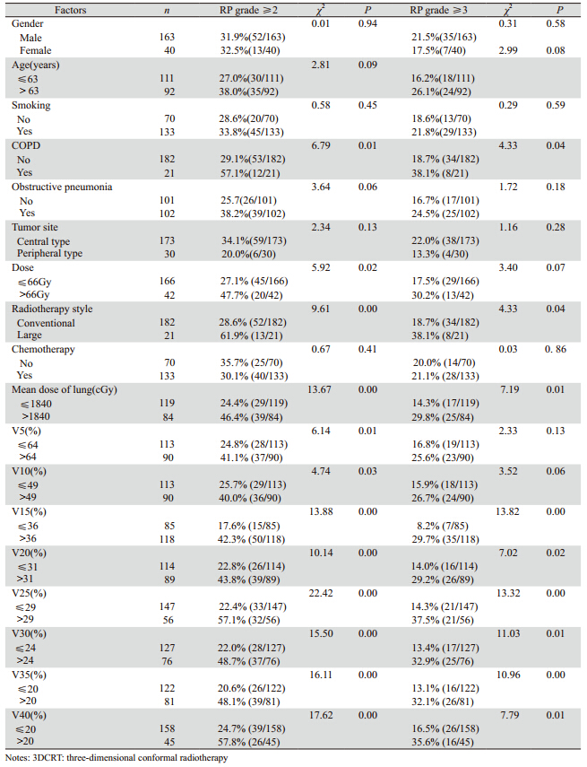
卡方检验显示,有CO P D病史、放疗剂量>66Gy、大分割照射者,≥2级和≥3级RP的发生率明显高于无COPD病史、放疗剂量≤66Gy、常规分割照射者(P<0.05);肺平均剂量、双肺V5~V40按照上述分界值分组均显示与≥2级和≥3级RP的发生率有关系,双肺受照剂量越高,RP的发生率越高(P<0.05);而性别、年龄、吸烟与否、原发瘤位置、化疗与否与≥2级和≥3级RP的发生率无明显差别(P>0.05),见表 3

表 3 Ⅲ期非小细胞肺癌患者三维适形放后放射性肺炎的影响因素 Table 3 Influence factors for RP in stage Ⅲ NSCLC patients after 3DCRT
3 讨论

放射性肺炎是Ⅲ期NSCLC放疗的主要剂量限制性因素之一。本研究发生≥2级急性期放射性肺炎(RP)者为32%(65/203);国外研究[1,2,3,4]报道,NSCLC放疗或放化疗后发生≥2级RP约为15%~31%;国内文献[5,6,7,8,9]报道,NSCLC单纯放疗或放化疗后≥2级RP发生率约为17%~37%,与本研究结果基本一致,提示预防或减少放射性肺炎是非常重要的。本研究中≥3级RP者发生率为20.7%(42/203)。国外研究报道[10,11,12,13,14],NSCLC放疗或放化疗后发生≥3级RP约为11%~32%,与本研究基本一致;国内文献报道[8,15,16]≥3级RP发生率约为9.5%~14%。NSCLC三维适形放疗或放化疗联合治疗时,应尽量避免出现≥3级RP,一旦发生会严重影响患者生存治疗,需积极治疗。

NSCLC三维适形放疗时,治疗计划系统提供的物理学参数不但可以评价肿瘤靶区的受照剂量,还可以较客观地评价肿瘤周围正常组织的剂量分布大小。本研究结果显示,双肺V5~V40、肺平均剂量(MLD)与≥2级RP和≥3级RP的发生均有关系,与国内外多数文献报道结果基本一致[2,5,6,7,8,9,17,18,19,20,21]。文献报道,NSCLC三维适形放疗或放化疗后,DVH提供的物理参数中,预测≥2级放射性肺炎的独立危险因素有:V5[6,18]、V20[3,8,9,18,20]、V30[2,4,18]、MLD[2,3,4,17,18,19,20]、NTCP[20]等;预测≥3级放射性肺炎的独立危险因素有V5[5,14,15]、V10[8,10,11] 、V20~V50[16]、MLD[12,16,19] 、NTCP[11]等,而本研究中显示≥2级和≥3级RP最有价值的预测因素为V25;考虑可能与病例组成、治疗方法、纳入统计的物理学指标不完全相同等因素有关;也提示单个因素用于预测放射性肺炎的价值有限。本研究中,ROC曲线提示V25预测≥2级和≥3级RP的敏感度和特异性均不够理想,可能需多个物理学因子综合考虑更要意义。本研究中利用ROC曲线计算得出了以下分界值:MLD(1 840 cGy)、V5(64%)、V10(49%)、V15(36%)、V20(31%)、V25(2 9 %)、V3 0(2 4 %)、V3 5(2 0 %)、V4 0(20%)。多个物理学参数综合评估,可以更好地预测放射性肺炎;同时也提示,在临床放疗实践中,应尽可能降低肺组织的受照剂量。

本研究中GTV、GTV最大剂量、GTV/肺体积越大,≥2级和≥3级RP发生率越高,反之RP发生率越低;而肺体积大小与≥2级和≥3级RP的发生无明显关系。Zhang等[18]报道,GTV大小与≥2级RP有关;Kong等[20]报道,肺体积、GTV大小与≥2级RP有关;Dang等[21]报道PTV/肺体积大小可预测≥2级RP,≥70岁老年患者放疗后出现≥3级和≥ 2级RP者,其PTV/肺体积要明显低于年轻患者。提示肿瘤靶体积大小和肿瘤/肺体积比值大小也影响RP,肿瘤体积大、肿瘤/肺体积比值越大者,制定放疗计划时,应尽可能降低肺的受照剂量,减少放射性肺炎的发生。

多数文献报道,放疗分割方式[3,7,13,14,15,22]和放疗剂量[2,3,5,7,9,12,13,14,15]大小与放射性肺炎的发生无明显关系。而本研究中,Ⅲ期NSCLC采用大分割照射者,≥2级和≥3级RP发生率明显高于采用常规分割者;另外放疗剂量大小也影响RP,剂量越高、≥2级和≥3级RP发生率越高,可能与本研究采用连续变量法统计、且本组中处方剂量差别较大(5 000~7 800 cGy)等因素有关。最近RTOG 0617研究[23]显示,高剂量照射未明显增加疗效,但不良反应明显增加,与本研究结果基本一致,提示Ⅲ期NSCLC三维适形放疗时放疗剂量不宜过高。

NSCLC三维适形放疗联合化疗是否增加RP发生率,文献报道结果不一致。Kim等[2]、Zhang等[18]报道,联合化疗者≥2级RP明显升高;Palma等[3]报道>65岁使用卡铂+紫杉化疗≥2级肺炎危险度>50%;Dang等[21]报道化疗是预测NSCLC放疗后是否发生≥3级RP的独立危险因素;Parashar等[22]报道,放化疗和单纯放疗相比RP发生率分别为62.7% (42/67) 和15.8% (3/19)(P< 0.001),放化疗联合RP发生率增加5倍;Dang等[19]报道放疗联合化疗者较单纯放疗者RP增加7.6倍。但也有文献报道[2,5,6,7,8,13,14],放化联合治疗与单纯放疗相比,并没有明显增加放射性肺炎的发生率;本研究中Ⅲ期NSCLC单纯三维适形放疗、序贯放化疗、同期放化疗比较,与≥2级和≥3级RP发生无明显差别,考虑可能与资料回顾性、化疗方案不统一、化疗强度不够、周期数偏少等因素有关。提示化疗是否增加放射性肺炎的发生率,可能与放疗剂量、化疗方案和剂量强度、患者一般状况、个体差异等诸多因素有关,需要综合考虑。

基础肺疾病是否增加放射性肺炎,文献报道结果不一。有文献报道[2,8,14],放疗前肺疾病或COPD与放射性肺炎之间无明显关系。本研究结果显示,合并COPD病史者≥2级和≥3级RP发生率明显增加,COPD是预测≥2级RP的独立危险因素;王静等[6]报道,合并基础肺疾病是≥2级RP的独立危险因素;Shi等[11]报道,COPD增加≥3级RP; Zhang等[18]采用Meta分析结果提示,基础肺疾病增加放射性肺炎的发生,与本研究结果基本一致。因此有COPD的Ⅲ期NSCLC制定放疗计划时应格外考虑肺的受照剂量,尽量减少放射性肺炎。

总之,Ⅲ期NSCLC三维适形放疗或放化疗后,放疗计划系统中提供的物理学参数可用于预测放射性肺炎的发生,双肺V25是预测≥2级和≥3级RP最有价值的物理指标,综合多个预测指标可能更有价值;另外合并COPD也是预测≥2级RP的重要危险因素。

参考文献
[1] Rodrigues G, Lock M, D’Souza D, et al. Prediction of radiation pneumonitis by dose-volume histogram parameters in lung cancer--a systematic review[J]. Radiother Oncol, 2004, 71(2):127-38.
[2] Kim M, LeeJ, Ha B, et al. Factors predicting radiation pneumonitis in locally advanced non-small cell lung cancer[J].Radiat Oncol J, 2011, 29(3): 181-90.
[3] Palma DA, Senan S, Tsujino K, et al. Predicting radiation pneumo nitis after chemoradiation therapy for lung cancer: an international individual patient data Meta-analysis[J].Int J Radiat Oncol Biol Phys, 2013, 85(2): 444-50.
[4] Fay M, Tan A, Fisher R, et al. Dose-volume histogram analysis as predictor of radiation pneumonitis in primary lung cancer patients treated with radiotherapy[J].Int J Radiat Oncol Biol Phys, 2005, 61(5): 1355-63.
[5] Fu HY, Lu B,Xu BQ, et al. Prospective study of lung V5 and V10 in predicting radiation-induced lung injury in advanced nonsmall-cell lung cancer treated with three-dimensional conformal radiation therapy[J]. Zhonghua Fang She Zhong Liu Xue Za Zhi, 2009, 18(6): 439-42. [付和谊, 卢冰, 徐冰清, 等. Ⅲ+Ⅳ期非小细胞肺癌三维适形放疗正常肺V5、V10预测放射性肺损伤前瞻性临床研究[J]. 中华放射肿瘤学杂志, 2009, 18(6): 439-42.]
[6] Wang J, Qiao XY, Cao YK, et al. Analysis of correlated factors of radiation pneumonitis after three-dimensional conformal radiotherapy for non-small cell lung cancer[J].Zhongguo Zhong Liu Lin Chuang, 2009, 36(19): 1086-9. [王静, 乔学英, 曹彦坤, 等. 非小细胞肺癌三维适形放疗放射性肺炎发生的多因素分析[J]. 中国肿瘤临床, 2009, 36(19): 1086-9.]
[7] Zhuang TT, Liu Q, Zhang L, et al. Risk factors of acute radiation pneumonitis in patients with non-small cell lung cancer after concurrent three-dimensional conformal radiotherapy and chemotherapy [J]. Zhonghua Fang She Zhong Liu Xue Za Zhi, 2009, 18(6): 443-7. [庄婷婷, 柳青, 张黎, 等. 非小细胞肺癌三维 适形放疗同期化疗后急性放射性肺炎发生因素分析[J].中华放射肿瘤学杂志, 2009, 18(6): 443-7.]
[8] Zhu XZ, Wang LH, Wang YJ, et al. Risk factors of radiation pneumonitis in stage III non-small cell lung cancer treated with three-dimensional conformal radiotherapy[J]. Zhonghua Fang She Zhong Liu Xue Za Zhi, 2007, 16(6): 421-8. [朱向帜, 王绿化, 王 颖杰, 等. 三维适形放疗局部晚期非小细胞肺癌的放射性肺炎 风险因素研究[J]. 中华放射肿瘤学杂志, 2007, 16(6): 421-8.]
[9] Wang DQ, Zhang J, Li BS, et al. Dose-volume histogram parameters for predicting radiation pneumonitis using receiver operating characteristic curve[J]. Zhonghua Fang She Yi Xue Yu Fang Hu Za Zhi, 2012, 32(5): 505-8. [王冬青, 张建, 李宝生, 等. 剂量体积直方图参数对放射性肺炎的工作特征曲线预测分析[J]. 中华放射医学与防护杂志, 2012, 32(5): 505-8.]
[10] Yom SS, Liao Z, Liu HH, et al. Initial evaluation of treatmentrelated pneumonitis in advanced-stage non-small-cell lung cancer patients treated with concurrent chemotherapy and intensity-modulated radiotherapy[J].Int J Radiat Oncol Biol Phys, 2007, 68(1): 94-102.
[11] Shi A, Zhu G, Wu H, et al. Analysis of clinical and dosimetric factors associated with severe acute radiationpneumonitis in patients with locally advanced non-small cell lung cancer treated with concurrent chemotherapy and intensity-modulated radiotherapy[J]. Radiat Oncol, 2010, 5: 35.
[12] Oh D, Ahn YC, Park HC, et al. Prediction of radiation pneumonitis following high-dose thoracic radiation therapy by 3 Gy/fraction for non-small cell lung cancer: analysis of clinical and dosimetric factors[J].Jpn J Clin Oncol, 2009, 39(3): 151-7.
[13] Jin H, Tucker SL, Liu HH, et al. Dose-volume thresholds and smoking status for the risk of treatment-relatedpneumonitis in inoperable non-small cell lung cancer treated with definitive radiotherapy [J].Radiother Oncol, 2009, 91(3): 427-32.
[14] Wang S, Liao Z, Wei X, et al. Analysis of clinical and dosimetric factors associated with treatment-relatedpneumonitis (TRP) in patients with non-small-cell lung cancer (NSCLC) treated with concurrent chemotherapy and three-dimensional conformal radiotherapy (3D-CRT)[J].Int J Radiat Oncol Biol Phys, 2006, 66(5): 1399-407.
[15] Wang J, Wang P, Pang QS, et al. Clinical and dosimetric factors associated with radiation-induced lung damage in patients with non small cell lung cancer treated with three-dimentional conformal radiotherapy[J]. Zhonghua Fang She Zhong Liu Xue Za Zhi, 2009, 18(6): 448-51. [王静, 王平, 庞青松, 等.非小细胞肺癌三维适形放疗放射性肺损伤临床及剂量学因素分析[J]. 中华放射肿瘤学杂志, 2009, 18(6): 448-51. ]
[16] Wang J, Zhang TT, He ZC, et al. Severe acute radiation pneumonitis after concurrent chemoradiotherapy in non-small cell lung cancer[J]. Zhonghua Fang She Zhong Liu Xue Za Zhi, 2012, 21(4): 326-9. [王谨, 庄婷婷, 何智纯, 等. 非小细胞肺癌同期放化疗后重度急性放射性肺炎的预测因素[J]. 中华放射肿瘤学杂志, 2012, 21(4): 326-9.]
[17] Wang D, Li B, Wang Z, et al. Functional dose-volume histograms for predicting radiation pneumonitis in locally advanced nonsmall cell lung cancer treated with late-course accelerated hyperfractionated radiotherapy[J].Exp Ther Med, 2011, 2(5): 1017-22.
[18] Zhang XJ, Sun JG, Sun J, et al. Prediction of radiation pneumon itis in lung cancer patients: a systematic review[J].J Cancer Res Clin Oncol, 2012, 138(12): 2103-16.
[19] Dang J, Li G, Lu X, et al. Analysis of related factors associated with radiation pneumonitis in patients with locally advanced nonsmall-cell lung cancer treated with three-dimensional conformal radiotherapy[J].J Cancer Res Clin Oncol, 2010, 136(8): 1169-78.
[20] Kong FM, Hayman JA, Griffith KA, et al. Final toxicity results of a radiation-dose escalation study in patients with non-smallcell lung cancer (NSCLC): predictors for radiation pneumonitis a nd fibrosis[J].Int J Radiat Oncol Biol Phys, 2006, 65(4): 1075-86.
[21] Dang J, Li G, Ma L, et al. Predictors of grade ≥ 2 and grade ≥ 3 radiation pneumonitis in patients with locally advanced nonsmall cell lung cancer treated with three-dimensional conformal radiotherapy [J].Acta Oncol, 2013, 52(6): 1175-80.
[22] Par ashar B, Edwar ds A, Mehta R, et al. Chemother apy significantly increases the risk of radiation pneumonitis in radiation therapy of advanced lung cancer[J].Am J Clin Oncol, 2011, 34(2): 160-4.
[23] Br adley JD, Paulus R, Komaki R, et al. A r andomized phase Ⅲ comparison of standard-dose(60Gy)versus highdose(74Gy)comformal chemoradio+/- cetuximab for stage III nonsmall cell lung cancer: results on radiation dose in RTOG 0617[J]. J Clin Oncol, 2013, 31(suppl;abstr 7501).